Congratulations on your achievement, glad you are keeping us up to date. Sounds like the UK and Australian exams are much harder than Canadian and American ones. I can't imagine how a solo consultant lab can provide quality "training" to a resident.
You are using an out of date browser. It may not display this or other websites correctly.
You should upgrade or use an alternative browser.
You should upgrade or use an alternative browser.
Disillusioned, need help staying motivated for AP
- Thread starter CDX-2
- Start date
If there was only one pathologist and one trainee in the lab, and the latter was mostly printing cassettes, our AP/CP exam surely would have a similar passing rate.Our AP/CP exam should be ~60% pass. Now, it is practically a gimme.
I felt like my AP/CP board was quite difficult. The passing “score” must be set super low to have the current passing rate.
- Joined
- Nov 16, 2014
- Messages
- 85
- Reaction score
- 52
That's because the questions are stupid. No one makes diagnoses based on one feature in one field of the slide without any clinical information whatsoever. Also, they had the opportunity to get a new set of slides scanned for future virtual microscopy exams but instead they scanned those old and decolorised Tampa slides and immortalized those bad slides forever. The stupidity of ABP and CAP knows no bounds...If there was only one pathologist and one trainee in the lab, and the latter was mostly printing cassettes, our AP/CP exam surely would have a similar passing rate.
I felt like my AP/CP board was quite difficult. The passing “score” must be set super low to have the current passing rate.
If there was only one pathologist and one trainee in the lab, and the latter was mostly printing cassettes, our AP/CP exam surely would have a similar passing rate.
I felt like my AP/CP board was quite difficult. The passing “score” must be set super low to have the current passing rate.
Yeah, we joke about US residents being scut monkeys, but if CDX-2's posts are even half-exaggerated the Australian training system sounds like total garbage. Congrats on making it through CDX-2!
Congratulations on your achievement, glad you are keeping us up to date. Sounds like the UK and Australian exams are much harder than Canadian and American ones. I can't imagine how a solo consultant lab can provide quality "training" to a resident.
Thank you shikimate, I'm so happy and relieved that I finally fully passed AP Part 1 (albeit it took me 5.5 years).
I have a 5 year time limit to pass AP Part 2 (Cytology / Small Biopsy / Slides / Viva), but hopefully it'll be less painful...
Hopefully I can fully complete AP training in Australia within 10 years or less, before the job market gets re-saturated / even more saturated...........?
RCPA (Australia / New Zealand / Singapore / Malaysia / Hong Kong + Saudi Arabia for some reason) slide exams require you to provide a written microscopic description and favoured diagnosis, or short list of DDx with a favoured Dx, along with relevant work-up (if required).
I think RCPath (UK) slide exams also require written answers.
I heard that the American AP slide exams are MCQ?!?!?!
In that sense, the RCPA slide exam is more difficult, purely due to the need to actually write stuff down (I wish it was MCQ
If it's a "spotter", you can get away with writing a much shorter microscopic description, and not have to list any DDx.
My personally submitted answers for this year's RCPA AP Part 1 slide exam (2022):
1. Favour Haemangioblastoma over met. clear cell RCC. Do IHC panel.
2. Paraganglioma / carotid body tumour. Do IHC.
3. Favour Pneumatosis Cystoides Intestinalis over Lymphangioma. Do D2-40 (expect negative).
4. Lipoid Pneumonia. Check if patient is swallowing castor oil.
5. Necrotizing lymphadenitis, favour Kikuchi's Disease. Do special Stains + PCR to exclude TB / fungi etc.
6. Favour non-invasive LGPUC.
7. Favour desmoid Fibromatosis. Do IHC panel including B-Catenin.
8. Favour Luteoma of Pregnancy over ovarian Leydig cell Tumour.
9. Benign Brenner's Tumour +/- ???Fibroma. Do Inhibin to check for Fibroma bits.
10. Favour goblet-cell rich hyperplastic polyp. DDx: Mucosal prolapse.
11. Giant cell tumour of bone + ?Aneurysmal bone cyst. Correlate with imaging.
12. Papillary breast lesion, favour intraductal Papilloma + focal UDH.
Need to do myoepithelial markers + ER to differentiate from encysted papillary Ca. and papillary DCIS.
13. Invasive tubular carcinoma of breast + DCIS (at least intermediate grade).
14. Sperm granulomas.
15. Endometriosis of the fallopian tube + ???Non-necrotizing granulomatous salpingitis. Do ER/CD10 and special stains for TB / fungi etc.
16. Chondroid syringoma.
17. Paediatric recurrent facial lesion. Benign spindle cell neoplasm, favour plexiform Neurofibroma.
18. LCH of mandible.
19. Hydatid cyst of liver.
20. Cystic adrenal lesion. I wrote adrenal cortical adenoma + [ ???cystic nephroma / ???benign mesothelial cyst / ???lymphangioma ].


NO OVARIAN-TYPE STROMA WAS SEEN IN THE CYST WALL.
Do Inhibin, Melan-A, ER, D2-40, Calretinin.
Apparently I scored @ least 16/20 correct this time.
APPARENTLY THIS IS MOSTLY A REFLECTION OF DAILY ROUTINE HISTOLOGY REPORTING IN AUSTRALIA??????????????????!!!!!!!!!!!!!!!!!!!!
Congratulations on your achievement, glad you are keeping us up to date. Sounds like the UK and Australian exams are much harder than Canadian and American ones. I can't imagine how a solo consultant lab can provide quality "training" to a resident.
I don't think they will.
Well, based on my experiences, I think it'd be very difficult for a solo consultant/attending to provide quality "training" for a solo registrar (resident), especially the buck's on them for histology / cytology / frozen sections / MDMs (multidisciplinary meetings, ie tumour boards).
It might work out if the solo registrar (resident) is very senior/experienced (which is not the case) and is capable of partially absorbing some of the solo consultant/attending's workload...
That original regional private lab where I got severely abused apparently had to advertize for 6 months to get that solo consultant/attending to work there.
And the only reason why they took the job is because they happen to have family there.
If that solo consultant/attending retires, I'll be curious to see how long they'll have to wait before the position gets filled again...............................
Last edited:
Yeah, we joke about US residents being scut monkeys, but if CDX-2's posts are even half-exaggerated the Australian training system sounds like total garbage. Congrats on making it through CDX-2!
I got off on a VERY BAD start for 1st and 2nd year AP in that original regional private lab.
VERY BAD doesn't even begin to describe it in retrospect.
NOT ALL LABS ARE LIKE THAT.
There are many GOOD labs in Australia where AP registrars (residents) engage in RELEVANT AP SERVICE PROVISION DUTIES (CUT-UP/GROSSING, AUTOPSIES, MEETINGS/TUMOUR BOARDS, HISTOLOGY REPORTING, CYTO ROSE),
and
not IRRELEVANT SCIENTIST SCUT WORK FOR MONTHS OR YEARS ON END (ACCESSIONING,
CASSETTE-PRINTING,
RE-LOADING THE CASSETTE PRINTER ONE CASSETTE AT A TIME,
HAND-WRITING CASSETTES WITH A PENCIL WHEN THE CASSETTE-PRINTER BREAKS DOWN,
VET CUT-UP/GROSSING,
DOING MANUAL DEFAT BY FILLING BUCKETS WITH ETOH + XYLENE AND THEN CHANGING THE FLUID EVERY 2 HOURS FOR THE BREAST AND BOWEL BLOCKS THAT YOU DID,
MS-DOS DATA ENTRY,
SPECIMEN THROWOUT,
SEARCHING AND SCANNING MISSING HISTO REQUEST FORMS THAT ARE HIDDEN AMONGST THE STACK OF SCANNED REQUEST FORMS).
I even got asked to do an AUTOPSY ON A DEAD PUPPY (??????????), and I said it was beyond my scope of practice!!!!!!!!!!!!!!!!!!!!!!!!!!!!!!!!!!!!!!!!!!!!!!!!!!!!!!!!!!!!!!
Unfortunately, in my original state where I started AP training, the ratio of GOOD labs to BAD labs seems to be lower than the other states..................
Hence the eventual "need" for me to move interstate to work in GOOD labs, where I'm doing RELEVANT AP REGISTRAR/RESIDENT WORK, and not being treated as a source of federally-funded free labour with no regards to educational or RELEVANT training requirements.
I think the company that owns the original regional private lab that I got abused in is starting to go down........................................
Last edited:
Yeah, we joke about US residents being scut monkeys, but if CDX-2's posts are even half-exaggerated the Australian training system sounds like total garbage. Congrats on making it through CDX-2!
Oh and thank you ScubaV!!!!!!!!!
- Joined
- Apr 3, 2007
- Messages
- 1,701
- Reaction score
- 722
What’s up with the 50 point size colored font?I got off on a VERY BAD start for 1st and 2nd year AP in that original regional private lab.
VERY BAD doesn't even begin to describe it in retrospect.
NOT ALL LABS ARE LIKE THAT.
There are many GOOD labs in Australia where AP registrars (residents) engage in RELEVANT AP SERVICE PROVISION DUTIES (CUT-UP/GROSSING, AUTOPSIES, MEETINGS/TUMOUR BOARDS, HISTOLOGY REPORTING, CYTO ROSE),
and
not IRRELEVANT SCIENTIST SCUT WORK FOR MONTHS OR YEARS ON END (ACCESSIONING,
CASSETTE-PRINTING,
RE-LOADING THE CASSETTE PRINTER ONE CASSETTE AT A TIME,
HAND-WRITING CASSETTES WITH A PENCIL WHEN THE CASSETTE-PRINTER BREAKS DOWN,
VET CUT-UP/GROSSING,
DOING MANUAL DEFAT BY FILLING BUCKETS WITH ETOH + XYLENE AND THEN CHANGING THE FLUID EVERY 2 HOURS FOR THE BREAST AND BOWEL BLOCKS THAT YOU DID,
MS-DOS DATA ENTRY,
SPECIMEN THROWOUT,
SEARCHING AND SCANNING MISSING HISTO REQUEST FORMS THAT ARE HIDDEN AMONGST THE STACK OF SCANNED REQUEST FORMS).
I even got asked to do an AUTOPSY ON A DEAD PUPPY (??????????), and I said it was beyond my scope of practice!!!!!!!!!!!!!!!!!!!!!!!!!!!!!!!!!!!!!!!!!!!!!!!!!!!!!!!!!!!!!!
Unfortunately, in my original state where I started AP training, the ratio of GOOD labs to BAD labs seems to be lower than the other states..................
Hence the eventual "need" for me to move interstate to work in GOOD labs, where I'm doing RELEVANT AP REGISTRAR/RESIDENT WORK, and not being treated as a source of federally-funded free labour with no regards to educational or RELEVANT training requirements.
I think the company that owns the original regional private lab that I got abused in is starting to go down........................................
- Joined
- Sep 8, 2019
- Messages
- 296
- Reaction score
- 733
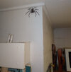
Probably helps deter the spiders.What’s up with the 50 point size colored font?
Attachments
- Joined
- Dec 16, 2010
- Messages
- 2,130
- Reaction score
- 1,032
Damn, I doubt my Mr Kitty would be killing that thing. Probably the other way around.
I think your slide exam feel more like the daily workload of a foreign trained slave at a "prestigious" academic center.
Of the 20 answers that I submitted for the 2022 RCPA AP Part 1 Digital Slide Exam,
I can say that I've personally only done draft histology reports on
#6 (Non-invasive LGPUC)
#7 (Desmoid Fibromatosis)
#10 (Goblet cell-rich hyperplastic Polyp)
#12 (Intraductal Papilloma + focal UDH)
#13 (Invasive tubular Carcinoma of Breast + DCIS)
#14 (Sperm Granulomas)
#15 (Endometriosis of the fallopian Tube)
#16 (Chondroid Syringoma)
As for my other submitted answers, I've only seen those entities in study slide sets, or via textbooks / internet.
I didn't realize that "prestigious" academic centres in the US had such esoteric pathology on a daily basis?!?!?!?!?!?!?!?!?!?!?!
The esoteric and difficult stuff usually comes from consults. The consults are often seen by clinical fellows. It's good for education to see these esoteric stuff but it's hard work and fellows aren't paid that much $ for seeing them.
I find the external consult part can take up a lot of consultant's energy and time as well. Now, if they charge a lot of $ and the consultant gets a good chunk, then there could be some motivation. But I've seen places whereby the consultants aren't given much for doing these difficult external consults and the department takes the majority chunk.
I find the external consult part can take up a lot of consultant's energy and time as well. Now, if they charge a lot of $ and the consultant gets a good chunk, then there could be some motivation. But I've seen places whereby the consultants aren't given much for doing these difficult external consults and the department takes the majority chunk.
The esoteric and difficult stuff usually comes from consults. The consults are often seen by clinical fellows. It's good for education to see these esoteric stuff but it's hard work and fellows aren't paid that much $ for seeing them.
I find the external consult part can take up a lot of consultant's energy and time as well. Now, if they charge a lot of $ and the consultant gets a good chunk, then there could be some motivation. But I've seen places whereby the consultants aren't given much for doing these difficult external consults and the department takes the majority chunk.
Oh I see.
In Australia, there are 2 billable items off Medicare for providing a second opinion / consult.
MBS (Medicare Benefits Schedule) Item #72858 - $AU 180 (if it took <=30 minutes to complete report)
MBS Item #72859 - $AU 370 (if it took more than 30 minutes to complete report)
In a public lab, the consultant (attending) pathologist gets paid as per the state/territory health sector EBA.
In the public and private labs I've worked in so far, most of the cases that I've seen that get referred to other labs tend to be T-cell lymphoproliferative neoplasms or unusual melanocytic lesions where it's not explicitly clear if it's malignant melanoma or a dysplastic/regressing naevus (even with IHC for SOX-10 / Melan-A / HMB-45 / PRAME).
Thanks for the info.
The consult rate varies greatly in Canada (I've seen anywhere from 50$ to >200$) but the consults sent to US are charged at a much higher rate, not to mention in USD (multiple hundreds). We only send to US if a clinician specifically request it or it's a very specialized testing that's not available in Canada.
The consult rate varies greatly in Canada (I've seen anywhere from 50$ to >200$) but the consults sent to US are charged at a much higher rate, not to mention in USD (multiple hundreds). We only send to US if a clinician specifically request it or it's a very specialized testing that's not available in Canada.
The official answers for the RCPA AP Part 1 slide exam (2022, digital) have been disclosed, for which I officially scored 18.5 / 20:
1. Haemangioblastoma
2. Paraganglioma
3. Hydatid cyst (liver)
4. Benign Brenner's tumour (ovary)
5. Intraductal papilloma (breast).
- Should provide IHC for myoepithelial markers.
6. Sperm granuloma
7. Adrenal cortical adenoma (had cystic change)
8. Necrotizing lymphadenitis.
- Required work-up to exclude TB/fungi.
- Needed to also list at least one of the following as DDx: Kikuchi, Lupus, Herpes, Cat scratch disease.
9. Pneumatosis intestinalis
10. Chondroblastoma
- I wrote giant cell tumour of bone + aneurysmal bone cyst --> Borderline mark (apparently lots of other people wrote this as well).
11. Chondroid syringoma
12. Non-invasive low-grade papillary urothelial carcinoma
13. Lipoid pneumonia
14. Luteoma (ovary)
15. Plexiform schwannoma
- I wrote plexiform neurofibroma --> Borderline mark.
16. Granulomatous salpingitis. Required work-up to exclude TB.
17. Langerhans cell histiocytosis (mandible)
18. Mucosal prolapse (needed to be listed as the favoured Dx)
- I favoured goblet-cell rich hyperplastic polyp but listed mucosal prolapse as DDx --> Borderline mark.
19. Desmoid fibromatosis. Needed to provide IHC work-up for bland spindle cell lesions.
20. Tubular carcinoma / Grade 1 invasive breast carcinoma + cribriform DCIS
Last edited:

