Ecmo coverage

This forum made possible through the generous support of SDN members, donors, and sponsors. Thank you.

anes121508

Full Member
10+ Year Member
Joined
Jan 16, 2012
Messages
745
Reaction score
293
hey all,

For those of you out there at places that do ecmo....

(I’ve noticed practice variations at different institutions)

is your anesthesia group providing coverage? If so, why?

Is tee routinely used for decannulation?Why or why not?


Where are the cannulas being put in? Icu or OR?

Where are thy being decannulated?

Members don't see this ad.
 
is your anesthesia group providing coverage? If so, why?

Is tee routinely used for decannulation?Why or why not?


Where are the cannulas being put in? Icu or OR?

Where are thy being decannulated?

Pediatric patients are put on ECMO in the ICU and there is no anesthesia involvement. They are also decannulated in the ICU, I'm assuming without TEE. Adult patients are frequently cardiac surgical patients that are more often than not put on ECMO in the OR during a related surgical procedure under anesthesia. It's hit or miss whether or not they are weaned from ECMO in the OR or in the ICU, but in the ICU there is no anesthesia involvement.
 
No anesthesia on the pager coverage, only surgeons and perfusionists go out for cannulation. They do it everywhere in the hospital, but if they decide to bring it to the OR it’s usually for a good reason and we will be present.

Decannulation depends on the cannulation method and type of ECMO. TEE is almost always used for VA decannulation and is done either in the ICU (peripheral) or the OR (central). VV is usually decannulated in the ICU without us present. If a cutdown was done for cannulation, they’ll usually come to the OR for decannulation regardless of the ECMO type.

Hope this helps!
 
  • Like
Reactions: 1 user
Members don't see this ad :)
No anesthesia on the pager coverage, only surgeons and perfusionists go out for cannulation. They do it everywhere in the hospital, but if they decide to bring it to the OR it’s usually for a good reason and we will be present.

Decannulation depends on the cannulation method and type of ECMO. TEE is almost always used for VA decannulation and is done either in the ICU (peripheral) or the OR (central). VV is usually decannulated in the ICU without us present. If a cutdown was done for cannulation, they’ll usually come to the OR for decannulation regardless of the ECMO type.

Hope this helps!

Thank you! This is probably the most straightforward organized response anyone has given me on this issue so far.

Sounds like decannulation (as opposed to cannulation) is where anesthesia may or may not be present.

I think there are two things to address.
1. Do you need an anesthetic?
2. Do you need a tee?

Then maybe: what if major bleeding happens?

Does this sound right?

decannulation TEE
1. VA ecmo

Decannulation OR:
1. VA placed via cutdown
2. VA percutaneously placed centrally

Decannulation ICU:
1. VA percutaneously placed peripherally
2. VV

Cannulation OR:
1. Cutdown needed

Cannulation icu:
1. Percutaneously
 
No anesthesia on the pager coverage, only surgeons and perfusionists go out for cannulation. They do it everywhere in the hospital, but if they decide to bring it to the OR it’s usually for a good reason and we will be present.

Decannulation depends on the cannulation method and type of ECMO. TEE is almost always used for VA decannulation and is done either in the ICU (peripheral) or the OR (central). VV is usually decannulated in the ICU without us present. If a cutdown was done for cannulation, they’ll usually come to the OR for decannulation regardless of the ECMO type.

Hope this helps!

To play devils advocate, is tee necesssary?

Can you decannulate without tee? Why or why not?

Why not tte as opposed to tee?
 
VV ECMO
-TEE to help position avalon in OR if stable enough to tolerate tx'fer
-femoral cannulation in ICU if unable to tolerate tx'fer
-almost always decannulate in ICU without anesthesia

VA ECMO
-usually cannulate in OR if possible
-almost always use TEE in OR for decannulation


VV ECMO is vey easy to know when you are ready to decannulate. Just "cap" the circuit and if they tolerate for a period of time remove the canulas.

VA ECMO decannulation is a more inexact science. Cant turn the flow all the way off. Higher incidence of crumping in the minutes to hours after decannulation. TEE very helpful here. Often the cardiologist is involved with these at my institution.
 
To play devils advocate, is tee necesssary?

Can you decannulate without tee? Why or why not?

Why not tte as opposed to tee?

VA ECMO placement is typically for cardiogenic shock or low output cardiac syndromes post CPB. IMO TEE is absolutely necessary for turndowns and decannulation because you need to look at the heart in real time as support is being weaned. This includes looking at how RV and LV fnx changes with the increased preload, evaluating worsening of any valvular abnormalities, inspecting the aorta, and looking for intracardiac clots and hemopericardium. The hemodynamics of VA ECMO are complex, and it's much easier to determine using TEE (if for instance MAP drops precipitously when you wean from 2L to 0L) whether it's due to hypovolemia or low SVR or primary LV failure or RV failure or wide open TR or MR due to annular distortions or impaired RA filling due to loculated clot etc etc etc. VA weaning is as much art as it is science and at the end of the day depends on integrating data from labs, CCO/SVO2 swan, TEE, and the pt's clinical condition.
 
  • Like
Reactions: 1 user
CV Anesthesiology and CT surgery both cannulate.
CV Anesthesia for VV, ARDS lung rescue with a call pool, VA, VAV for eCPR in-hospital arrest
CT surgery for ED ECMO (rare), OR cases (Obviously), If CT surgery is not operating then they will be around for hands on help for VV, etc.
Perfusion runs machines.
Pre-lung transplant and intraop lines for transplant are still being worked out with CT surgery and CV Anesthesia. The general tone is CT surgeons are more friendly in the sandbox with CV Anesthesia then Cardiology. All Devices (LVADs, ECMO, Centrimag, RVAD, IABP) go to CT ICU that is pure Anesthesia and CT surgery. Obviously MIs with impella, IABP stay in CCU.

TEE for all cannulation. Usually there is an IMACOR hTEE left for 48 hours. Rarely the patient will need flouro, which if needed can be done at bedside with a C ARM in the unit.

Most decannulation is in the ICU without "anesthesia" - but not sure what that refers to since the CT ICU is anesthesia staff and likely they had input and decision making into the timing of decannulation.
If OR decannulation then anesthesia is doing "anesthesia."

The overall theme is devices, ICU care, and cannulation is time and labor intensive. It can be financially beneficial for a CV group and usually the CV surgeons are comfortable with Anesthesia staff. Start with TEE, then ICU care, and then cannulation and rescue.


Super helpful reply thank you!

So your decannulation is occurring in either the OR or the ICU? What determines the location? Some have argued that decannulation only should occur in the OR.

Also noticed you brought up the imacor hTEE being used continuously after cannulation, however when you discussed decannulation you didn’t mention echo. I’m assuming tee is involved?
 
VV ECMO
-TEE to help position avalon in OR if stable enough to tolerate tx'fer
-femoral cannulation in ICU if unable to tolerate tx'fer
-almost always decannulate in ICU without anesthesia

VA ECMO
-usually cannulate in OR if possible
-almost always use TEE in OR for decannulation


VV ECMO is vey easy to know when you are ready to decannulate. Just "cap" the circuit and if they tolerate for a period of time remove the canulas.

VA ECMO decannulation is a more inexact science. Cant turn the flow all the way off. Higher incidence of crumping in the minutes to hours after decannulation. TEE very helpful here. Often the cardiologist is involved with these at my institution.

So your decannulations are almost always done in the OR? Why OR vs ctu?

Also, you mentioned crumping in min to hours after turning flow down. Are you doing a trial in the ctu with lower flows and serial tte over the course of a few hours, and then going to the OR to decannulate? How long are you staying in the OR with tee monitoring before you decide to head back to the ctu?
 
VV ECMO
-TEE to help position avalon in OR if stable enough to tolerate tx'fer
-femoral cannulation in ICU if unable to tolerate tx'fer
-almost always decannulate in ICU without anesthesia

VA ECMO
-usually cannulate in OR if possible
-almost always use TEE in OR for decannulation


VV ECMO is vey easy to know when you are ready to decannulate. Just "cap" the circuit and if they tolerate for a period of time remove the canulas.

VA ECMO decannulation is a more inexact science. Cant turn the flow all the way off. Higher incidence of crumping in the minutes to hours after decannulation. TEE very helpful here. Often the cardiologist is involved with these at my institution.


Also interesting that you brought up cardiology being involved during decannulation in the OR. They are doing the tee?
 
Reason I post the original question is because we are in the process of trying to figure it out. Seems there are different methods at different places with no universal approach....

However what I gather to be true are a few things (please correct me where I am wrong, very open to suggestions and new points)

1. Support devices are labor and time intensive
- financially beneficial for the cv group?

2. Echo is an invaluable tool to evaluate ability to wean off ecmo
- not necessarily a quick tee but rather a continual evaluation over the course of 30min - 2hrs or however long pt is at risk for crumping?

3. Places with ecmo programs have a anesthesia staff involved with the icu and are part of the patients management team
- anyone out there like us, where anesthesia has zero presence in the ctu yet we are being asked to come in for tee evaluation at time of decannulation?
 
Can anyone share their ecmo weaning process or protocol?

Are you using impellas in combination with the ecmo to help transition?

Anyone using a bridge on the ecmo cannulas and clamping off? (So the patient is excluded from the circuit and the ecmo can run without clotting)

Either way Seems to me like it’s a process that is being continually evaluated by the intensivists that are most often tee capable. almost like vent weaning. Underlying problem improving, minimal pressors/inotropes, wean down flows on ecmo and monitor hemodynamics and serial tte or tee evaluations to look at the heart over the course of hours...then decide to decannulate. Location tbd. Accurate?

Today I had a case:

Called to do tee in OR last min (wasn’t covering the room, just asked to help). Asked for the story on the patient. Surgeon getting ready to decannulate peripheral va ecmo has no clue. Perfusionist has no clue. Impella in place, can’t remember settings but low. Ecmo at full flow. Dobuta running. On tee the lv is completely shot ef maybe 10% and enormous. Rv working. Impella in good position. No clots anywhere. I get cardiology on the phone because not sure what’s the plan here. Cards (who staffs the ctu), says they turned the flows down to 2 yesterday for 30 min and pt hemodynamics remained stable. Today was day 10 and last day they wanted him on ecmo. Cocaine intox. Not transplant or vad candidate. Just pull and bring him back to CTU with impella and whatever gtts we needed.

Had them turn down the ecmo flow while surgeon worked in groin. Added some milrinone and watched the tee. Lv couldn’t get any worse, rv stayed the same. 30 min later cannulas were out and we went back to CTU.

Comments? I Just feel a bit of disconnect since I’m not totally part of the management of these patients and I’m seeing them at a snap shot in time. Also trying to figure out my role since we aren’t involved in the ctu at all. Seems like we may be doing something wrong here? Suggestions?
 
Members don't see this ad :)
Can anyone share their ecmo weaning process or protocol?

Are you using impellas in combination with the ecmo to help transition?

Anyone using a bridge on the ecmo cannulas and clamping off? (So the patient is excluded from the circuit and the ecmo can run without clotting)

Either way Seems to me like it’s a process that is being continually evaluated by the intensivists that are most often tee capable. almost like vent weaning. Underlying problem improving, minimal pressors/inotropes, wean down flows on ecmo and monitor hemodynamics and serial tte or tee evaluations to look at the heart over the course of hours...then decide to decannulate. Location tbd. Accurate?

Today I had a case:

Called to do tee in OR last min (wasn’t covering the room, just asked to help). Asked for the story on the patient. Surgeon getting ready to decannulate peripheral va ecmo has no clue. Perfusionist has no clue. Impella in place, can’t remember settings but low. Ecmo at full flow. Dobuta running. On tee the lv is completely shot ef maybe 10% and enormous. Rv working. Impella in good position. No clots anywhere. I get cardiology on the phone because not sure what’s the plan here. Cards (who staffs the ctu), says they turned the flows down to 2 yesterday for 30 min and pt hemodynamics remained stable. Today was day 10 and last day they wanted him on ecmo. Cocaine intox. Not transplant or vad candidate. Just pull and bring him back to CTU with impella and whatever gtts we needed.

Had them turn down the ecmo flow while surgeon worked in groin. Added some milrinone and watched the tee. Lv couldn’t get any worse, rv stayed the same. 30 min later cannulas were out and we went back to CTU.

Comments? I Just feel a bit of disconnect since I’m not totally part of the management of these patients and I’m seeing them at a snap shot in time. Also trying to figure out my role since we aren’t involved in the ctu at all. Seems like we may be doing something wrong here? Suggestions?

That's a bizarre story. Never heard of anyone getting a flyby call to the OR to TEE a VA ecmo decannulation- where I've been these have always been booked with CT anesthesia. Since cards runs CTICU there, I'm surprised at the very least a cardiologist wasn't booked to TEE the decannulation.

Very quickly, this sounded like a possible trainwreck of a case, and I have no idea why the impella was set to a low setting. Had the impella been at low flows for awhile? The whole point of impella + ECMO is to decompress the LV, get the inotropes down a bit, and allow the LV to recover from a distension and energy use standpoint. If you walk into the room and the LV is totally blown up and the EF is 10% then it sounds more like a terminal decannulation than a successful wean.

If it was me, I would've seen if the impella flow rate could've been turned up as your bridge to decannulation since the RV looked good. Usually you'll get limited bang for your buck trying to flog the LV with more inotropes, but if the patient made it out of the OR then I guess it was at least somewhat a success. I'll typically go epi + milrinone + iNO (prn) for these pts. Two inodilators vis a vis dobu+mil might be a bit hairy from a MAP standpoint once the ECMO is off. Did you have PAC in place and/or shoot a doppler CO? What did the hemodynamics look like as you were leaving the room?
 
Can anyone share their ecmo weaning process or protocol?

Are you using impellas in combination with the ecmo to help transition?

Anyone using a bridge on the ecmo cannulas and clamping off? (So the patient is excluded from the circuit and the ecmo can run without clotting)

Where I did residency, we had one surgeon who did it like this. My sense was that this was the more conservative way to decannulate.


Today I had a case:

Called to do tee in OR last min (wasn’t covering the room, just asked to help). Asked for the story on the patient. Surgeon getting ready to decannulate peripheral va ecmo has no clue. Perfusionist has no clue. Impella in place, can’t remember settings but low. Ecmo at full flow. Dobuta running. On tee the lv is completely shot ef maybe 10% and enormous. Rv working. Impella in good position. No clots anywhere. I get cardiology on the phone because not sure what’s the plan here. Cards (who staffs the ctu), says they turned the flows down to 2 yesterday for 30 min and pt hemodynamics remained stable. Today was day 10 and last day they wanted him on ecmo. Cocaine intox. Not transplant or vad candidate. Just pull and bring him back to CTU with impella and whatever gtts we needed.

Had them turn down the ecmo flow while surgeon worked in groin. Added some milrinone and watched the tee. Lv couldn’t get any worse, rv stayed the same. 30 min later cannulas were out and we went back to CTU.

Comments? I Just feel a bit of disconnect since I’m not totally part of the management of these patients and I’m seeing them at a snap shot in time. Also trying to figure out my role since we aren’t involved in the ctu at all. Seems like we may be doing something wrong here? Suggestions?

What size Impella was it? If the RV was fine and it was an Impella 5.0, the ECMO may have been overkill to begin with.
 
  • Like
Reactions: 1 user
hey all,

For those of you out there at places that do ecmo....

(I’ve noticed practice variations at different institutions)

is your anesthesia group providing coverage? If so, why?

Is tee routinely used for decannulation?Why or why not?


Where are the cannulas being put in? Icu or OR?

Where are thy being decannulated?
Me during this whole thread::corny:
 
  • Like
Reactions: 1 user
Thrilled I don’t have to be on call for this sort of stuff. They just grab someone else who’s available - our institution isn’t too keen on ECMO’ing everyone under the sun (as they did in fellowship) as it’s a pretty expensive prospect and often the patients don’t do all that well.
 
That's a bizarre story. Never heard of anyone getting a flyby call to the OR to TEE a VA ecmo decannulation- where I've been these have always been booked with CT anesthesia. Since cards runs CTICU there, I'm surprised at the very least a cardiologist wasn't booked to TEE the decannulation.

Very quickly, this sounded like a possible trainwreck of a case, and I have no idea why the impella was set to a low setting. Had the impella been at low flows for awhile? The whole point of impella + ECMO is to decompress the LV, get the inotropes down a bit, and allow the LV to recover from a distension and energy use standpoint. If you walk into the room and the LV is totally blown up and the EF is 10% then it sounds more like a terminal decannulation than a successful wean.

If it was me, I would've seen if the impella flow rate could've been turned up as your bridge to decannulation since the RV looked good. Usually you'll get limited bang for your buck trying to flog the LV with more inotropes, but if the patient made it out of the OR then I guess it was at least somewhat a success. I'll typically go epi + milrinone + iNO (prn) for these pts. Two inodilators vis a vis dobu+mil might be a bit hairy from a MAP standpoint once the ECMO is off. Did you have PAC in place and/or shoot a doppler CO? What did the hemodynamics look like as you were leaving the room?

I think you bring up good points. Forgot to mention that I turned up the impella at same time of decreasing ecmo flows from the get go. Figured this would have been done before getting to the OR. So I agree with your approach to these.

We had a pa catheter in place and the numbers didn’t really budge with turning the flows down and upping the impella. Didn’t have cco capabaility. Manually shot the output and can’t quote numbers but I was surprised that the CI looked decent. I shot some pwd in the lvot since I’ve read that some people are using vti as a metric, but I’m unsure if you could really use this with an impella in place. What are your thoughts on pwd of lvot and sv calculation in setting of impella?
 
Where I did residency, we had one surgeon who did it like this. My sense was that this was the more conservative way to decannulate.


What size Impella was it? If the RV was fine and it was an Impella 5.0, the ECMO may have been overkill to begin with.

It was a 5.0. Can you elaborate on this? I think I get what you are saying and make a good point.
 
Me during this whole thread::corny:

It’s is fairly entertaining because I’m not sure that this a well ironed out topic. No really best way to do it and tons of variation. Take that combined with this being fairly new at my place and it makes for some interesting ....”wait what are we doing? Why is this patient here? What’s the plan? Why are the settings the way they are? What’s the end game?”
 
It was a 5.0. Can you elaborate on this? I think I get what you are saying and make a good point.

If the RV and lungs were fine, and the guy's only problem was a weak LV, it's possible that 5 liters per minute of cardiac output through the Impella was enough. I don't know what the sequence of MCS devices was (i.e. maybe he got the VA ECMO emergently, and then had the Impella placed in the cath lab during daytime hours?) but it sounds like he maybe could've gotten away with just the Impella.
 
I think you bring up good points. Forgot to mention that I turned up the impella at same time of decreasing ecmo flows from the get go. Figured this would have been done before getting to the OR. So I agree with your approach to these.

We had a pa catheter in place and the numbers didn’t really budge with turning the flows down and upping the impella. Didn’t have cco capabaility. Manually shot the output and can’t quote numbers but I was surprised that the CI looked decent. I shot some pwd in the lvot since I’ve read that some people are using vti as a metric, but I’m unsure if you could really use this with an impella in place. What are your thoughts on pwd of lvot and sv calculation in setting of impella?

I was also under the impression that you can't measure VTI in the LVOT with an Impella in place. Is this incorrect? We honestly don't use many Impellas in my shop for our surgical patients. Mostly just used by our interventional Cardiologists
 
  • Like
Reactions: 1 user
I think you bring up good points. Forgot to mention that I turned up the impella at same time of decreasing ecmo flows from the get go. Figured this would have been done before getting to the OR. So I agree with your approach to these.

We had a pa catheter in place and the numbers didn’t really budge with turning the flows down and upping the impella. Didn’t have cco capabaility. Manually shot the output and can’t quote numbers but I was surprised that the CI looked decent. I shot some pwd in the lvot since I’ve read that some people are using vti as a metric, but I’m unsure if you could really use this with an impella in place. What are your thoughts on pwd of lvot and sv calculation in setting of impella?

Sorry, had a brainfart and totally forgot about the impella when asking about doppler. The mixture of continuous + pulsatile flow in the LVOT would presumably make a VTI based CO estimate inaccurate, but I doubt anyone has studied it.
 
Also, wanted to share an excellent, excellent presentation about mechanical circulatory support. The guy is a cardiologist and has done a lot of work with pressure/volume loops and the different devices out there. There's also some info on the "pulmonary artery pulsatility index" which increasingly looks like a good way to figure out which pts will need BiV support.

The biggest takeaway point is that various devices differ significantly in how much they actually rest the ventricle. VA-ECMO is circulatory support, not ventricular support, and LV stroke work actually goes up when VA-ECMO is initiated.

 
Last edited:
If the RV and lungs were fine, and the guy's only problem was a weak LV, it's possible that 5 liters per minute of cardiac output through the Impella was enough. I don't know what the sequence of MCS devices was (i.e. maybe he got the VA ECMO emergently, and then had the Impella placed in the cath lab during daytime hours?) but it sounds like he maybe could've gotten away with just the Impella.

You are correct. That’s exactly what happened.
 
Also, wanted to share an excellent, excellent presentation about mechanical circulatory support. The guy is a cardiologist and has done a lot of work with pressure/volume loops and the different devices out there. There's also some info on the "pulmonary artery pulsatility index" which increasingly looks like a good way to figure out which pts will need BiV support.

The biggest takeaway point is that various devices different significantly in how much they actually rest the ventricle. VA-ECMO is circulatory support, not ventricular support, and LV stroke work actually goes up when VA-ECMO is initiated.


Great discussion. Thank you for posting this. Haven’t found great resources....

If va ecmo is increasing lv stroke work then that’s the reasoning for the impella combo?
 
Great discussion. Thank you for posting this. Haven’t found great resources....

If va ecmo is increasing lv stroke work then that’s the reasoning for the impella combo?

More accurate is probably to say that the stroke work is about the same. It may be somewhat higher or lower depending on the MAP after VA ECMO is initiated.

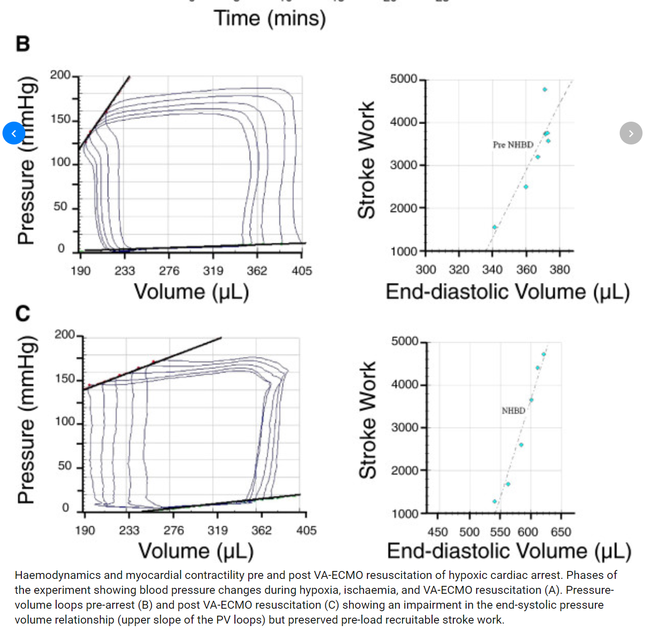
Take a look at the this diagram. It's a rat study (pre-arrest measurements, hypoxic arrest, initiation of VA-ECMO), but the priniciples are the same:

1YfOsOY.png


You can see between the pre loops on the top left and the post loops on the bottom left that the area under the curve (stroke work) isn't significantly different once VA ECMO is initiated. The "afterload" sensed by the ventricle is still very high compared to an impella where you have active LV decompression. Ultimately with MCS you want a PV loop that looks like a slurred triangle, not a rectangle. I'm definitely of the opinion that any pt whose ventricle is bad enough for VA ECMO needs an impella or other LV vent strategy while the ventricle recovers.
 
  • Like
Reactions: 1 user
More accurate is probably to say that the stroke work is about the same. It may be somewhat higher or lower depending on the MAP after VA ECMO is initiated.

Take a look at the this diagram. It's a rat study (pre-arrest measurements, hypoxic arrest, initiation of VA-ECMO), but the priniciples are the same:

1YfOsOY.png


You can see between the pre loops on the top left and the post loops on the bottom left that the area under the curve (stroke work) isn't significantly different once VA ECMO is initiated. The "afterload" sensed by the ventricle is still very high compared to an impella where you have active LV decompression. Ultimately with MCS you want a PV loop that looks like a slurred triangle, not a rectangle. I'm definitely of the opinion that any pt whose ventricle is bad enough for VA ECMO needs an impella or other LV vent strategy while the ventricle recovers.

Agree with above. I am pushing for our group CV Intensivists/Anesthesiologists who cannulate for ECMO to learn and implement Impella placement. Some of the folks from the Cards group here have offered to help guide us in impella placement. Work in progress. Will report back in ~ 1 year with our outcomes.
 
Agree with above. I am pushing for our group CV Intensivists/Anesthesiologists who cannulate for ECMO to learn and implement Impella placement. Some of the folks from the Cards group here have offered to help guide us in impella placement. Work in progress. Will report back in ~ 1 year with our outcomes.

Your anesthesiologists cannulate for ECMO? That is pretty cool.
 
Agree with above. I am pushing for our group CV Intensivists/Anesthesiologists who cannulate for ECMO to learn and implement Impella placement. Some of the folks from the Cards group here have offered to help guide us in impella placement. Work in progress. Will report back in ~ 1 year with our outcomes.

Interesting. What’s the story behind how you all trained for ecmo cannulation? My understanding is that this is not the norm for an icu fellowship.
 
Trained at Michigan? I've heard their fellows do that a lot. We scrubbed in to assist the CT surgeon (often one do venous, the other arterial and retrograde perfusion) when we cannulated in the unit during my fellowship, but it was still largely surgeon driven.

Sent from my SM-G930V using SDN mobile
 
We have a local community hospital where the pulm/icu guy cannulates V-v ecmo. There is no CT surgery program there.
 
Interesting. What’s the story behind how you all trained for ecmo cannulation? My understanding is that this is not the norm for an icu fellowship.

To my knowledge Michigan, Vandy, Emory, Penn, UCSD and others are open to training ICU + CV folks. It's in the pipelines for sure. The thought being you dont need a cardiologist to deploy an iabp, impella, or ecmo in a crashing pt. In the middle of the night
 
Only going to post my experiences with V-V ECMO which is what I do most often. We do it in the unit, but have cardiac surgery + perfusion involved with placement. Anesthesia doesn't get involved, but sometimes we will call the anesthesiologist covering TEE for the day to assist with confirming cannula placement, though if I'm on or other intensivists who can do TEE we will just do it ourselves.
 
Top