Let's do some echo:

This forum made possible through the generous support of SDN members, donors, and sponsors. Thank you.
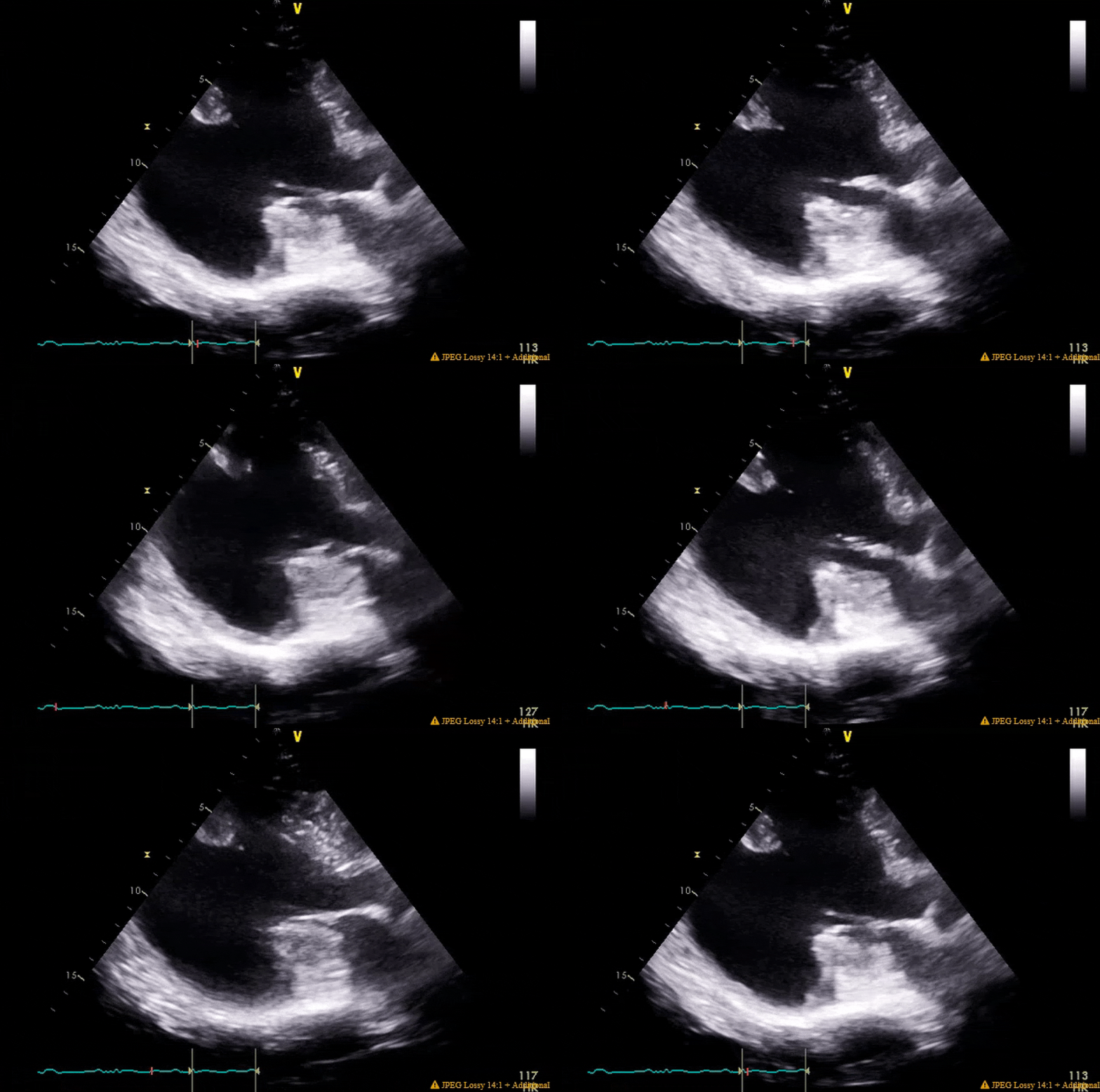
Pretty much spot on. The gifs didn't capture the way I would've liked (they're kinda choppy), but if you squint real hard you'll notice in the 4ch color shot that there is a central jet and a highly, eccentric anteriorly directed jet. There ended up being a cleft plus myxomatous degeneration leading to both Carpentier type I and II mechanisms.

You'll also notice the LV function is pretty bad pre-pump, and absolutely atrocious post-pump. The RV function is also moderately to severely depressed post-pump. Both the clamp time and pump run were very short, MVR went without a hitch. I expressed to my resident that this case highlighted the difference between doing pump cases for revascularization in ischemic cardiomyopathy with viable myocardium vs doing valvular surgery for people with non-ischemic cardiomyopathy. In the literature, AMI cardiogenic shock does better than decompensated NICM cardiogenic shock, and I find that to be generally (and anecdotally) true in the OR as well when one has post-cardiotomy shock.

The post-pump LV clip with an EF of ~15% is with the pt on high dose epi, dobutamine, milrinone, iNO, levo, vaso and IABP with a barely acceptable MAP and CI. He went on to require a takeback to the cathlab a few hours after he arrived in the ICU for impella placement due to worsening shock, and that highlighted some of the system issues we face with having surgeons who do not routinely put in their own impellas, VA-ECMO, LV vents, etc. They tried to punt on this case (either for transfer or possible clip), but pressure from cards was heavy given this was a young man (about age 50) with good functional status.

Finally, these clips highlight that in some patients you are just not going to be able to get a classic valentine heart 4 chamber view. I tried every combination of omniplane and flexion but his cardiomegaly and rotation made it impossible get a clip where I wasn't severely obliquely cutting through the LA. People btch a lot about the quality of clips in the aPTE, but difficult views are not terribly infrequent IRL.


Patient needed a VAD not a mitral intervention. Big hearts with **** function getting on pumps to fix a mitral lesion is a good case of attempted murder. The fact that he was 50 is even more criminal.

Members don't see this ad.
 
Patient needed a VAD not a mitral intervention. Big hearts with **** function getting on pumps to fix a mitral lesion is a good case of attempted murder. The fact that he was 50 is even more criminal.

He needed a mitral valve intervention. He's 50 with symptomatic structural mitral valve disease.

Also, at baseline he didn't have NYHA IV symptoms, an ef under 25 (although arguable in the setting of severe MR), inotrope dependence, nor do I think he was on all the latest greatest GDMT drugs.

What he did need though was to have his surgery done at the advanced HF/VAD/tx center across town, but I'm not sure if his cardiologists at my resource-poor institution told him that was an option, and/or whether his insurance is accepted there.
 
Last edited:
  • Like
Reactions: 2 users
Patient needed a VAD not a mitral intervention. Big hearts with **** function getting on pumps to fix a mitral lesion is a good case of attempted murder. The fact that he was 50 is even more criminal.
What?

A vad for severe MR?
 
Members don't see this ad :)
RCARWMA.gif


Question for residents (and maybe fellows but might be too easy):

1. What coronary distribution typically supplies the wall with the regional wall motion abnormality?

2. How do you explain it if I told you an angiogram of the coronary that typically supplies that wall is normal?
Not in training yet but will take a stab. Thanks for doing these.
1) Inferior wall motion abnormalities which is typically supplied by RCA->PDA
2) The patients PDA must be a branch of the circumflex which is why it doesn’t show up on the angiogram.
 
Not in training yet but will take a stab. Thanks for doing these.
1) Inferior wall motion abnormalities which is typically supplied by RCA->PDA
2) The patients PDA must be a branch of the circumflex which is why it doesn’t show up on the angiogram.
Very good, nailed it.

And very unfortunate for the patient in question. She had significant LM, proximal LAD, proximal Cx, and PDA lesions. Due to her left dominance, almost all her LV was at risk. Which wouldn't have been the case had she been right dominant like the majority of people, since her RCA was surprisingly pretty clean.
 
  • Like
Reactions: 1 users
What?

A vad for severe MR?
Severe MR and cardiomyopathy, yes. EF on that one pre-TEE looks 20-25ish, young guy, would be a relatively straightforward intermacs 4 VAD. I trained at a very aggressive VAD/txp center (>50 txps, >100 VADs) and this guy would have definitely been worked up at least, probably would have gone down the BTT VAD route. NYHA III by ROADMAP trial with drastic QOL improvement.
 
Severe MR and cardiomyopathy, yes. EF on that one pre-TEE looks 20-25ish, young guy, would be a relatively straightforward intermacs 4 VAD. I trained at a very aggressive VAD/txp center (>50 txps, >100 VADs) and this guy would have definitely been worked up at least, probably would have gone down the BTT VAD route. NYHA III by ROADMAP trial with drastic QOL improvement.
Good for you guys. No way this guy gets a primary vad before addressing his mitral in any other centre possibly in the world.

What is it a bridge to? Dt? Transplant? Bridge to MVR?
 
Good for you guys. No way this guy gets a primary vad before addressing his mitral in any other centre possibly in the world.

What is it a bridge to? Dt? Transplant? Bridge to MVR?
Should have got an axillary impella at time of mitral surgery then slow wean in ICU over next few days.
 
  • Like
Reactions: 2 users
Figured I'd revive this thread for a bit and give everyone a much-needed relief from the politics that have been dominating a lot of the forum --

Had some profound hypotension during a non-cardiac case last month. It's a pathology I'm aware of and this patient population is prone to it, but the speed of the decompensation surprised me -- went from 120s/60s to 50s/30s in ~2 minutes despite some volume and rocket fuel during the nosedive. Once we figured it out and treated it appropriately she did fine.

This one is probably way too easy for the staff around, but if there are any residents/fellows who want to answer --

a) what's the pathology?
b) what case / patient population makes this pathology more likely?
c) how would you treat this patient?


1680524714098.png


1680524448840.png
 
Last edited:
  • Like
Reactions: 3 users
Sam with MR and obstruction from hocm; fluids, vasoconstrictors (eg vasopressin, phenylepi), beta blockers. Need to avoid contractility and low afterload due to worsening the obstruction.
 
  • Like
Reactions: 1 user
Sam with MR and obstruction from hocm; fluids, vasoconstrictors (eg vasopressin, phenylepi), beta blockers. Need to avoid contractility and low afterload due to worsening the obstruction.

Correct! SAM with MR and obstruction. No HOCM, though - but she does have a chronic pathology that typically causes low SVR and thus can predispose to an LVOT gradient.

She was given ~1L volume quickly with CVP 11>22 with minimal improvement in BP. Would you continue the volume challenge? LV still looks underfilled, RV not dilated but TR has increased from tr > mild/mod and IVC looks distended.

Would you be comfortable giving her a beta blocker with a BP of ~50s/30s, or would you wait until her BP improves with your other interventions?
Ya beta blockers is a hard sell to someone in shock, but could try esmolol after using phenylepi to up her Svr. Could also try some inhaled nitric if no pulm edema to help fill the left, but obv wean quickly if sats drop or she develops pulm edema.

A swan here would be helpful to titrate these drips and to help establish hemos.
 
Correct! SAM with MR and obstruction. No HOCM, though - but she does have a chronic pathology that typically causes low SVR and thus can predispose to an LVOT gradient.

She was given ~1L volume quickly with CVP 11>22 with minimal improvement in BP. Would you continue the volume challenge? LV still looks underfilled, RV not dilated but TR has increased from tr > mild/mod and IVC looks distended.

Would you be comfortable giving her a beta blocker with a BP of ~50s/30s, or would you wait until her BP improves with your other interventions?

Are you saying no HOCM because they lacked that formal diagnose prior to this surgery/echo? Or are you saying that because there were features present on imaging that you felt weren’t consistent with HOCM? Because by my read you cinched the diagnosis of HOCM with your exam. They have clear asymmetric septal hypertrophy that looks >1.5cm (if it’s not 1.5 it’s wicked close) on cursory glance, as well as clear evidence of obstruction (flow acceleration in LVOT) with SAM.
 
  • Like
Reactions: 2 users
Members don't see this ad :)
She was given ~1L volume quickly with CVP 11>22 with minimal improvement in BP. Would you continue the volume challenge? LV still looks underfilled, RV not dilated but TR has increased from tr > mild/mod and IVC looks distended.

Would you be comfortable giving her a beta blocker with a BP of ~50s/30s, or would you wait until her BP improves with your other interventions?
LV needs time to fill. Turn off all your inotropes (mentioned you were on jet fuel), if tachycardic I would give 30-50mg esmolol, chase with 500-1000mcg neo, with those hemos and that echo, follow with a unit of vaso if hemos still in toilet. Get neo and vaso in line and infusing if they aren’t already.
 
Are you saying no HOCM because they lacked that formal diagnose prior to this surgery/echo? Or are you saying that because there were features present on imaging that you felt weren’t consistent with HOCM? Because by my read you cinched the diagnosis of HOCM with your exam. They have clear asymmetric septal hypertrophy that looks >1.5cm (if it’s not 1.5 it’s wicked close) on cursory glance, as well as clear evidence of obstruction (flow acceleration in LVOT) with SAM.

Full disclosure that I am not a CTA trained person.

I am saying that because after I consulted cardiology postop and reviewed the images with them, they told me the patient did not meet criteria. She also had a totally normal TTE, though they did not measure the upper septum that I saw (nor did I, and my remote image viewing software won't allow me to. Probably have to do that from the office.) They commented "The left ventricular cavity size and wall thickness are normal." on her TTE, and it specifically was written for ?SAM. They also had the not-particularly-shocking finding on her postop TTE off pressors and well resuscitated she had no evidence of SAM.

Reviewing the diagnostic criteria, "The diagnosis of HCM is confirmed with the presence of a left ventricular wall thickness of ≥15 mm that is otherwise unexplained by abnormal loading conditions (e.g., hypertension, valvular, congenital disease)." We seem to see a not-insignificant number of patients (~5%?) presenting for OLTx who have some evidence of DLOTO with dobutamine stress but none at rest, and my understand is cards has said this is remodeling from their chronic low SVR / high CO state but does not represent true HOCM.

Curious to hear the hive mind's thoughts - I did some Googling and it's not obvious from the diagnostic criteria whether she would qualify or not (seems like it depends on whether you consider her cirrhosis to be a contributing etiology, to my read). https://www.asecho.org/wp-content/u...-of-Patient-w-Hypertrophic-Cardiomyopathy.pdf


As far as the treatment--

I appreciate those of you who are brave enough to push beta blockers in this case with a BP that low. I wasn't willing until I got her SVR up. She got ~4 milligrams of neo, 10 units of vaso, and 1.5mg/kg of methylene blue over the course of 10 minutes and finally turned the corner. Once we got her SVR up she did very well and was ultimately almost off vasoactives at the end of the case.

I get what you (and by proxy the cardiologist) are saying here. I just think it’s getting into a really dangerous game of semantics. They have hypertrophied myocardium resulting in dynamic LVOT obstruction. That is irrefutable based on imaging and hemodynamics.

138B5E11-436F-4BD1-8353-443B3103A89A.jpeg


You can see the 1cm lines along the right side. No way that septum is less than 15mm in the LVOT. Their TTE just wasn't catching the LVOT at the same angles you were on TEE. If we aren’t calling that HOCM because it’s not genetically inherited, fine, then let’s call it non-inherited hypertrophied myocardium with dynamic LVOT onstruction. NIHMWDLVOTO for short…

Whether it’s genetically inherited or secondary to remodeling from a chronic low SVR state sounds like exactly the type of thing a cardiologist would pontificate on. It also sounds like the type of thing that will get this patient killed during their next anesthetic.

Some anesthesiologist will read that TTE report and cardiology note, be falsely reassured, blast them with 200 of prop and it will be a clean kill, just like it almost was for you.
 
  • Like
  • Haha
Reactions: 4 users
Full disclosure that I am not a CTA trained person.

I am saying that because after I consulted cardiology postop and reviewed the images with them, they told me the patient did not meet criteria. She also had a totally normal TTE, though they did not measure the upper septum that I saw (nor did I, and my remote image viewing software won't allow me to. Probably have to do that from the office.) They commented "The left ventricular cavity size and wall thickness are normal." on her TTE, and it specifically was written for ?SAM. They also had the not-particularly-shocking finding on her postop TTE off pressors and well resuscitated she had no evidence of SAM.

Reviewing the diagnostic criteria, "The diagnosis of HCM is confirmed with the presence of a left ventricular wall thickness of ≥15 mm that is otherwise unexplained by abnormal loading conditions (e.g., hypertension, valvular, congenital disease)." We seem to see a not-insignificant number of patients (~5%?) presenting for OLTx who have some evidence of DLOTO with dobutamine stress but none at rest, and my understand is cards has said this is remodeling from their chronic low SVR / high CO state but does not represent true HOCM.

Curious to hear the hive mind's thoughts - I did some Googling and it's not obvious from the diagnostic criteria whether she would qualify or not (seems like it depends on whether you consider her cirrhosis to be a contributing etiology, to my read). https://www.asecho.org/wp-content/u...-of-Patient-w-Hypertrophic-Cardiomyopathy.pdf


As far as the treatment--

I appreciate those of you who are brave enough to push beta blockers in this case with a BP that low. I wasn't willing until I got her SVR up. She got ~4 milligrams of neo, 10 units of vaso, and 1.5mg/kg of methylene blue over the course of 10 minutes and finally turned the corner. Once we got her SVR up she did very well and was ultimately almost off vasoactives at the end of the case.
I would call this ASH/DUST/HOCM with SAM and LVOTO, and I would further ask cards to do provocative maneuvers and imaging in their echo lab.
 
  • Like
Reactions: 1 user
I get what you (and by proxy the cardiologist) are saying here. I just think it’s getting into a really dangerous game of semantics. They have hypertrophied myocardium resulting in dynamic LVOT obstruction. That is irrefutable based on imaging and hemodynamics.

View attachment 368835

You can see the 1cm lines along the right side. No way that septum is less than 15mm in the LVOT. Their TTE just wasn't catching the LVOT at the same angles you were on TEE. If we aren’t calling that HOCM because it’s not genetically inherited, fine, then let’s call it non-inherited hypertrophied myocardium with dynamic LVOT onstruction. NIHMWDLVOTO for short…

Whether it’s genetically inherited or secondary to remodeling from a chronic low SVR state sounds like exactly the type of thing a cardiologist would pontificate on. It also sounds like the type of thing that will get this patient killed during their next anesthetic.

Some anesthesiologist will read that TTE report and cardiology note, be falsely reassured, blast them with 200 of prop and it will be a clean kill, just like it almost was for you.

Thanks for the input.

I wasn't trying to make the semantic argument myself so much as perhaps trying to parse the guidelines to understand why I got the answer I did from the CV consult service at my shop. I found them to be (unfortunately, typically - at least at my institution) dismissive of what had happened intraoperatively. They didn't even look at my images initially until I prompted them to do so but relied solely on the TTE images, which I thought was unfortunate. Among the many reasons I don't tend to consult cardiology for "preoperative clearance" unless I have a very specific question, because I've found them to be unhelpful (and perhaps ignorant) of the insults that occur in the OR.

I do wonder what the appropriate follow-up plan for this patient would be. With the exception of the extreme situation of a liver txp, she has tolerated many anesthetics and two dobutamine stress echos in the past without any evidence of DLVOTO or SAM. That said, she clearly has a predisposition that in certain circumstances can lead to severe hemodynamic instability.

Her TEE post improvement in her SVR:

1680561880279.png


So here's a question: would you have called cardiology back and forced them to come up with a follow-up plan for this patient? It seems like most of the risk of sudden cardiac death is related to genetic HOCM variants that can precipitate arrhythmias. It's challenging from the standpoint of consulting cardiology, explaining my concerns and then essentially getting blown off by the consult attending.
 
Thanks for the input.

I wasn't trying to make the semantic argument myself so much as perhaps trying to parse the guidelines to understand why I got the answer I did from the CV consult service at my shop. I found them to be (unfortunately, typically - at least at my institution) dismissive of what had happened intraoperatively. They didn't even look at my images initially until I prompted them to do so but relied solely on the TTE images, which I thought was unfortunate. Among the many reasons I don't tend to consult cardiology for "preoperative clearance" unless I have a very specific question, because I've found them to be unhelpful (and perhaps ignorant) of the insults that occur in the OR.

I do wonder what the appropriate follow-up plan for this patient would be. With the exception of the extreme situation of a liver txp, she has tolerated many anesthetics and two dobutamine stress echos in the past without any evidence of DLVOTO or SAM. That said, she clearly has a predisposition that in certain circumstances can lead to severe hemodynamic instability.

Her TEE post improvement in her SVR:

View attachment 368859


So here's a question: would you have called cardiology back and forced them to come up with a follow-up plan for this patient? It seems like most of the risk of sudden cardiac death is related to genetic HOCM variants that can precipitate arrhythmias. It's challenging from the standpoint of consulting cardiology, explaining my concerns and then essentially getting blown off by the consult attending.

Q, did you or anyone else get CW Doppler gradients through the LVOT/mid LV cavity during the episode?
 
Thanks for the input.

I wasn't trying to make the semantic argument myself so much as perhaps trying to parse the guidelines to understand why I got the answer I did from the CV consult service at my shop. I found them to be (unfortunately, typically - at least at my institution) dismissive of what had happened intraoperatively. They didn't even look at my images initially until I prompted them to do so but relied solely on the TTE images, which I thought was unfortunate. Among the many reasons I don't tend to consult cardiology for "preoperative clearance" unless I have a very specific question, because I've found them to be unhelpful (and perhaps ignorant) of the insults that occur in the OR.

I do wonder what the appropriate follow-up plan for this patient would be. With the exception of the extreme situation of a liver txp, she has tolerated many anesthetics and two dobutamine stress echos in the past without any evidence of DLVOTO or SAM. That said, she clearly has a predisposition that in certain circumstances can lead to severe hemodynamic instability.

Her TEE post improvement in her SVR:

View attachment 368859


So here's a question: would you have called cardiology back and forced them to come up with a follow-up plan for this patient? It seems like most of the risk of sudden cardiac death is related to genetic HOCM variants that can precipitate arrhythmias. It's challenging from the standpoint of consulting cardiology, explaining my concerns and then essentially getting blown off by the consult attending.
Great question. I’m echo boarded so I would have a signed echo report in the chart to accompany these images along with appropriate measurements and gradients (if I had time, which is a big if in situations like this). That report would read something to the tune of asymmetric septal hypertrophy, measuring Xmm at the widest dimension, with dynamic LVOT obstruction and systolic anterior motion of the mitral valve with associated hemodynamic collapse (insert vitals at time of collapse) consistent with HOCM.

Yes most of the risk for sudden cardiac death 2/2 HOCM is arythmogemic however if this patient ever gets bad gastro, a bowel prep, NPO for any serious period of time, vigorous activity with insensible losses and tachycardia, those are all life potentially threatening prospects for this patient that deserve proper consideration and attention. If I felt the experience in the OR was being written off as something other than what it was, and that the pathology in question was being ignored or not taken seriously, I would probably send a message to the cardiologist asking them to reconsider and take the patients ASH/LVOTO/SAM more seriously.
 
Lol who is giving beta blockers to someone with a BP of 50. Bad idea no matter the physiology

There are plenty of patients with regular old LVH from oldness/hypertension that develop SAM. This is not true HOCM as far as I understand (the genetic condition). But as you said just as prone to badness with an Anaesthetic
 
Quickie for residents and fellows as I was reviewing this cardiology echo.

What's the TR Vmax in the following?

1687348363729.png
 
  • Like
Reactions: 1 user
Quickie for residents and fellows as I was reviewing this cardiology echo.

What's the TR Vmax in the following?

View attachment 373298
Is this definitely the TR jet? And not accidentally catching some AS? Or maybe some LVOT to RA flow? Hard to tell with only that small pic up top.

Either way, I'd think that I would mark the peak of the whiter/denser jet, giving you TR Vmax closer to the 4-4.5m/s. I don't know the right answer here. Never been confronted with this challenge before.
 
Here's a 2d CFD clip of what they're insonating

bxIQjtj.gif
Given the velocities I'm inclined to think maybe some sort of gerbode defect. I think I'm seeing a teeny hint of L->R flow just below the septal tricuspid leaflet.

Or this guy's got some crazy pulm HTN.
 
Given the velocities I'm inclined to think maybe some sort of gerbode defect. I think I'm seeing a teeny hint of L->R flow just below the septal tricuspid leaflet.

Or this guy's got some crazy pulm HTN.

Since it’s on the ventricular side, there would have to be a septal leaflet perforation, typically congenital but can be iatrogenic, for it to be considered a gerbode defect, which there may be, I just can’t appreciate it on the images provided. I see the L>R flow you’re mentioning though. In the absence of a leaflet perforation, congenital or otherwise, just looks like a very high VSD. Definitely contributing to TR and right sided pressures though. Love the relatively obscure echo dx!
 
  • Like
Reactions: 1 user
Guess it's been long enough I'll just spill the beans.

As far as etiology, there is a hint as to causation in the clip. What's going on with the L side of the heart?

In the clip what's apparent on the L side is the appearance of 1. At least high moderate to severe MAC, 2. Flow acceleration / turbulence in the small part of the color box that includes mitral inflow. When we see obvious R sided pathology, first thing we should do is look for obvious left sided pathology before moving on to other zebras.


Another follow up question: if I tell you the true TR jet PV is 4.5 m/s and the IVC is >2.5 cm with less than 50% inspiratory variability, what's' this pt's RVSP?

Everyone learns about estimating RVSP/PASP from TR jets and estimating RAP from the IVC collapsibility index, but a point that is often overlooked is that the echocardiographic assessment of RAP simply falls apart in the face of severe TR and severe RA dilatation.

Firstly, just calling a plethoric IVC with no collapsibility in the pt above "15" is not going to be accurate. It could be 15. But it could also be 25. Or 35. And secondly, that goes along with the fact that TR jet velocity significantly goes down as the coaptation gap becomes massive and the RA enlarges. It's not unusual for regurgitant velocities to be less than 2 m/s when the TR is torrential (almost like common chamber physiology). So, if you decide to calculate the PASP as 16+15, odds are you're gonna be doing some pretty significant underestimation.
 
  • Like
Reactions: 1 users
IqNyR87.gif


What modality / view is this?

What are we looking at? Describe the wall motion.

What are the indications for surgical repair?
 
Last edited:
  • Like
Reactions: 1 user
IqNyR87.gif


What modality / view is this?

What are we looking at? Describe the wall motion.

What are the indications for surgical repair?
Looks like tilted PLAX on TTE
Is that an LVAD cannula with sucking in of the septal wall?
LV looks big with poor motion of the rest of the LV wall.
And there’s what looks like a big thrombus on the PMVL.
 
IqNyR87.gif


What modality / view is this?

What are we looking at? Describe the wall motion.

What are the indications for surgical repair?

LAX on epicardial ultrasound.

Big pseudo aneurysm of the inferior/infero-lateral wall.

Indication: you know it when you see it but in seriousness, I say this is worth fixing.
 
  • Like
Reactions: 1 users
LAX on epicardial ultrasound.

Big pseudo aneurysm of the inferior/infero-lateral wall.

Indication: you know it when you see it but in seriousness, I say this is worth fixing.

How do you make the diagnosis of pseudoaneurysm vs aneurysm?
 
IqNyR87.gif


What modality / view is this?

What are we looking at? Describe the wall motion.

What are the indications for surgical repair?
Big inferior wall aneurysm.

Refractory chf, VT, and I believe size are all indications for surgical intervention.
 
  • Like
Reactions: 1 user
How do you make the diagnosis of pseudoaneurysm vs aneurysm?
Generally, neck of pseudoaneurysm is less than 1/2 the diameter of the outpouching. Also, aneurysm is full thickness myocardium while pseudoaneurysm may vary from part of the myocardium to only pericardium containing it.

And to add, I believe you should always fix a pseudoaneurysm, as it's basically a contained free-wall rupture.
 
Generally, neck of pseudoaneurysm is less than 1/2 the diameter of the outpouching. Also, aneurysm is full thickness myocardium while pseudoaneurysm may vary from part of the myocardium to only pericardium containing it.

Indeed, if you're just eyeballing the clip then you might think the cavity is like 2x the neck so this must be a pseudo, but that's kinda a general rule, not a law.

Regardless of ratio, usually when you see a massive neck like in this example it's just a regular aneurysm. Additionally, the echo texture of the aneurysm wall looks like really thin, smooth infarcted endocardium/myocardium (which it is), whereas a pseudo is a contained rupture and the only thing holding it in is a heterogeneous mishmash of myocardium, clot and pericardium.

Also, this is just a TTE PLAX (with either the probe rocked or a rib space down), not epicardial.

Big inferior wall aneurysm.

Refractory chf, VT, and I believe size are all indications for surgical intervention.

Correct.

Legit q: any idea what size threshold is worthy of surgical consideration? Up-to-dated it the other day and there were no numbers. Didn't do a lit search tho
 
Last edited:
Looks like tilted PLAX on TTE
Is that an LVAD cannula with sucking in of the septal wall?
LV looks big with poor motion of the rest of the LV wall.
And there’s what looks like a big thrombus on the PMVL.
Lol way off
 
IqNyR87.gif


What modality / view is this?

What are we looking at? Describe the wall motion.

What are the indications for surgical repair?

Immediate gut response was pseudo based entirely on neck. But this clip does a great job of demonstrating how patients don’t read the textbooks. On closer look there’s definitely some thinned myocardium.

But if it’s infarcted, why is it moving? Asking the audience, not @vector2.
 
Different view and a clip with contrast

HeqMSQ4.gif

S0Wj7A2.gif


Notice how big the neck looks here and that the aneurysmal portion totally opacifies
 
  • Like
Reactions: 1 user
Top