ECG waves and cardiac action potentials are complicated topics that are covered in detail in medical school. I don't know what the passage says but I can provide a general overview. A good source to follow:
Physiology of cardiac conduction and contractility | McMaster Pathophysiology Review
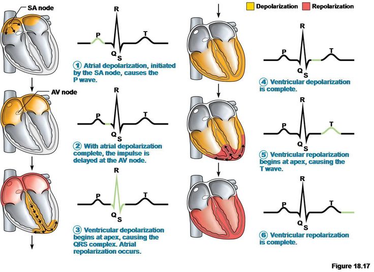
So take a look at the following diagram illustrating the relationship between ECG waves and cardiac depolarization/repolarization.
The key takeaways are the following:
P waves represent atrial depolarization
QRS complex represents ventricular depolarization and atrial repolarization
T waves represent ventricular repolarization
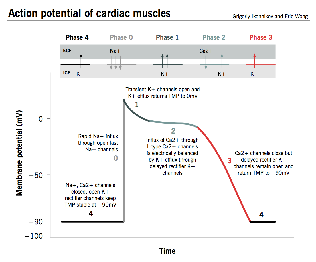
Under normal conditions, sodium and calcium have significantly higher extracellular concentrations, while potassium had significantly higher intracellular concentratios. So during depolarization, sodium and calcium flow into the cell, whereas in repolarization, potassium flows out of the cell. At the end of the cardiac action potential, normal ion concentrations are restored by sodium-calcium exchangers, calcium pumps, and sodium-potassium pumps.
The resting cardiac potential is -90 mV due to a steady outward leak of potassium through inward rectifier channels. Sodium and calcium channels are closed. The P wave occurs because of atrial depolarization that starts from the sinoatrial node, which serves as a cardiac pacemaker. The steps involved in the pacemaker potential are the initial flow of ions through slow sodium channels (called funny or pacemaker current) that increases the cardiac potential to above -60 mV, followed by opening of T-type calcium channels to continue slow depolarization. -40 mV is the threshold potential for pacemaker cells, where L-type calcium channels activate and quickly overshoots to +40 mV. Delayed rectifier potassium channels open to repolarize the pacemaker cells.
After atrial depolarization, the impulse is delayed at the atrioventricular node (this delay is measured by PR segment). The QRS complex represents ventricular depolarization while the atrial repolarization occurs roughly at the same time. The steps for ventricular depolarization are similar to atrial depolarization except here fast sodium channels are involved and are rapidly activated after -70 mV. L-type calcium channels open around -40 mV and continue the depolarization. Some potassium channels open while fast sodium channels close, reducing the overshoot to 0 mV. Calcium channels remain open as potassium channels begin to open, so calcium inflow matches potassium outflow to create a plateau. As calcium channels begin to close, potassium outflow is steady resulting in repolarization.
So in both atrial and ventricular depolarizations, sodium and calcium inflows are involved. And in both atrial and ventricular repolarizations, potassium outflow is involved. The following charts also serve as useful references:


