Question on MHC Molecules

This forum made possible through the generous support of SDN members, donors, and sponsors. Thank you.

CrimsonMirage

Full Member
10+ Year Member
15+ Year Member
Joined
Aug 15, 2008
Messages
106
Reaction score
0
Hi,

I'm a little confused about how MHC I vs MHC II molecules are actually made and what the differences in their final structures is. I just got a sample test question asking about invariant chain, beta-2 microglobulin, etc. Please help!

Thanks.

Members don't see this ad.
 
Hi,

I'm a little confused about how MHC I vs MHC II molecules are actually made and what the differences in their final structures is. I just got a sample test question asking about invariant chain, beta-2 microglobulin, etc. Please help!

Thanks.
Hmmm...maybe post the question?

MHC Class I= alpha chain + B2 micro globulin. (Loading in rough ER)

MHC Class II= alpha chain + beta chain (+ invariant chain before antigen loading). (Loading in Endosomes)

Invariant chain is associated with MHC Class II. Invariant chain needs to be removed in order for the antigen to bind to MHC Class II in endosomes. So it is only transiently a part of the molecule.
 
Hey there,

It's important to know the following things for MHC I:
  • Examples include HLA-A, HLA-B, and HLA-C
  • Expressed on ALL nucleated cells
  • Antigen is loaded in the rough endoplasmic reticulum
  • Largely involved in viral immunity
  • beta-2 microglobulin is a part of the structure

And then it's important to know the following for MHC II:
  • Examples include HLA-DR, HLA-DP, HLA-DQ
  • Expressed ONLY on the surface of antigen-presenting cells
  • Antigen is loaded following the release of the invariant chain

Hope that helps?
 
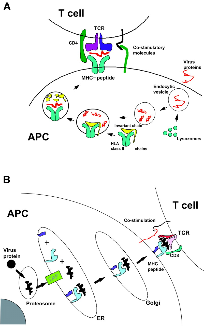
Picture time.

F1.large.jpg


Note: MHC-I are on ~all nucleated cells. MHC-II are on APC's, for presentation of Exogenous (phagocytosed) antigens. Small blue squiggly in the MHC-I diagram is the b2 microglobulin, found on a different gene (chromosome 15) compared to the rest of the MHC-I (chromosome 6).
 
Members don't see this ad :)
Picture time.

F1.large.jpg


Note: MHC-I are on ~all nucleated cells. MHC-II are on APC's, for presentation of Exogenous (phagocytosed) antigens. Small blue squiggly in the MHC-I diagram is the b2 microglobulin, found on a different gene (chromosome 15) compared to the rest of the MHC-I (chromosome 6).

Haha cool photos. Did you make these? Just of note, MHC Class II is a little bit less likely to display viral antigens. That's mainly a Class I thing.
 
The following is extra information that I had annotated into my FA after having done some USMLE Rx question a while ago (which I had invariably gotten wrong). I can't entirely recall what the specific question was, but this is apparently what I had felt necessary to annotate (iow, I'm prepared for this type of WTF question, if it happens to me):

MHC-I:

- SEVERAL alpha-helices & beta-2-globulin
- presented peptide binds in a groove in the ALPHA-helix.

MHC-II:

- TWO alpha & TWO beta-helices
- presented peptide binds in a groove formed by BOTH alpha- and beta-helices.
 
Wow, thanks everyone! That really cleared up my MHC confusion. And a special thanks for those pictures :)
 
Top