TheMysteryMan
New Member
Advertisement - Members don't see this ad
Disclaimer
I am a student of electrical engineering, not medicine. I have come here to bounce my idea off of those who are educated in dermatology. Please excuse my medical ignorance.
Introduction
I have an idea for a minimally invasive method for removing raised skin-colored benign moles/abnormalities that I'd like to bounce off of some of you dermatologists/students.
Background
I was looking around online for surgical techniques for dealing with raised skin-colored moles and I found that there were no methods that resulted in minimal scarring. In addition, there were no surgical instruments that could accommodate the procedure that I had in mind.
Thesis Statement
I believe it may be possible to achieve minimal scarring in the removal of benign raised skin-colored abnormalities by boring into the epidermis/dermis with a coaxial suction-saw.
Topic 1: The Tool
I believe I can develop and manufacture a tool that could internally excise and remove benign tissue. The tool would resemble a needle with a nominal outside diameter of no more than 0.9 mm (20 gauge needle) with a wall thickness of no more than 0.3 mm. The internal 0.6 mm space would contain coaxial angled razors that would rotate through radial grooves in the needle wall towards the tip of the needle. The outside diameter of the internal rotating shaft connected to the razors would be 0.4 mm. The remaining 0.2 mm tolerance would be used as the vacuum. The rear of the needle tool could be connected to a vacuum and this would serve to evacuate excised tissue.
Topic 2: Procedure Aftermath
After a sufficient amount of benign tissue has been excised from the epidermis/dermis, a compression bandage would be applied to the area. This would render the previously raised area flush with the surrounding skin. I believe the development of granulation tissue and epithelial cells would continue as normal considering the depth of the procedure. The resulting scar would be circular with a diameter of 0.9 mm. This is a vast improvement over ordinary excision techniques which would result in a linear scar with a length that sometimes exceeds 6 mm (depending on the size of the skin abnormality).
Conclusion
Do you suppose this is a reasonable procedure? What complications might arise from taking such an approach to excising benign tissue as opposed to conventional methods?
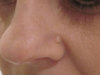
Note
The image included shows an example of a benign raised skin-colored abnormality that would be a candidate for this procedure.
I am a student of electrical engineering, not medicine. I have come here to bounce my idea off of those who are educated in dermatology. Please excuse my medical ignorance.
Introduction
I have an idea for a minimally invasive method for removing raised skin-colored benign moles/abnormalities that I'd like to bounce off of some of you dermatologists/students.
Background
I was looking around online for surgical techniques for dealing with raised skin-colored moles and I found that there were no methods that resulted in minimal scarring. In addition, there were no surgical instruments that could accommodate the procedure that I had in mind.
Thesis Statement
I believe it may be possible to achieve minimal scarring in the removal of benign raised skin-colored abnormalities by boring into the epidermis/dermis with a coaxial suction-saw.
Topic 1: The Tool
I believe I can develop and manufacture a tool that could internally excise and remove benign tissue. The tool would resemble a needle with a nominal outside diameter of no more than 0.9 mm (20 gauge needle) with a wall thickness of no more than 0.3 mm. The internal 0.6 mm space would contain coaxial angled razors that would rotate through radial grooves in the needle wall towards the tip of the needle. The outside diameter of the internal rotating shaft connected to the razors would be 0.4 mm. The remaining 0.2 mm tolerance would be used as the vacuum. The rear of the needle tool could be connected to a vacuum and this would serve to evacuate excised tissue.
Topic 2: Procedure Aftermath
After a sufficient amount of benign tissue has been excised from the epidermis/dermis, a compression bandage would be applied to the area. This would render the previously raised area flush with the surrounding skin. I believe the development of granulation tissue and epithelial cells would continue as normal considering the depth of the procedure. The resulting scar would be circular with a diameter of 0.9 mm. This is a vast improvement over ordinary excision techniques which would result in a linear scar with a length that sometimes exceeds 6 mm (depending on the size of the skin abnormality).
Conclusion
Do you suppose this is a reasonable procedure? What complications might arise from taking such an approach to excising benign tissue as opposed to conventional methods?
Note
The image included shows an example of a benign raised skin-colored abnormality that would be a candidate for this procedure.