It's an anatomy explanation, but I can't piece it together. Would anybody mind helping me out with the answer to this?
Thanks!
Thanks!

It's an anatomy explanation, but I can't piece it together. Would anybody mind helping me out with the answer to this?
Thanks!
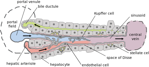
TREAT with splenectomy -> watch out for encapsulated bacteria in the futureNormally, portal blood drains to liver where the absorbed nutrients are processed and bugs are squashed by kupffer cells..
In cirrhosis there is increased resistance to portal blood flow due to fibrosis.
The increasing back pressure causes dilation of normally insignificant porto-caval shunts. Portal blood has to drain somewhere, right?
It is particularly more pronounced (and clinically significant) at the lower 1/3rd of esophagus since they are more prone to rupture due to submucosal nature of the dilated veins.
Since part of the portal blood is drained via spleen, esophageal varices can also be caused by splenic vein thrombosis (uncommon).
From Pathoma:
There are 2 ways by which veins of the esophagus drain: most of the esophageal blood drains via the azygous vein into the superior vena cava, but some of the blood drains from the left gastric vein into the portal vein - so some of the blood from the esophagus does drain into the portal circuit.
If there's portal hypertension --> you'll have a backup of blood into the left gastric vein, which will cause backup of blood into the veins of the esophagus that were draining via this pathway --> it results in dilation of those veins = Esophageal varices (dilated submucosal veins in the lower esophagus).
