Difficult Airway - Covid, Angioedema, ETT Exchange

This forum made possible through the generous support of SDN members, donors, and sponsors. Thank you.
Right, as in, if the paO2 doesnt require 100% fi02 , why are they prone at all? I would think that I would reserve turning prone (and all the complications that go with it) for those who have no other option, they are already maxed out on other oxygenation efforts..

can you provide evidence that it reduces mortality?

From what I have seen the evidence of reduced mortality is marginal even in a study with questionable methods

Here is an interesting one: Prone position for acute respiratory failure in adults. - PubMed - NCBI

And also, Im pretty sure that If i tried as hard as the guys who do most ICU studies, I could eek out a study that showed marginal benefit to recruitment manuevers and bronchodilators... or almost any intervention that i chose to study

I cant tell you how many times I have a patient with respiratory distress, no asthma history (but just had an LMA or a ETT in place) low sats, someone says "they are not wheezing" you give albuterol and they feel better and sats improve..

I think beta agonists are being underused from what Im seeing for that exact reasoning. You don't have to necessarily have a history of asthma or acute bronchospasm to get some oxygenation benefit from albuterol, especially when you have a viral infection of your airway causing some degree of reactivity.

All fair points. You could argue that inhaled beta agonists are acting as inhaled pulmonary vasodilators.

I think that I see plenty of improvement in people with proning and that I wouldn’t want to wait till they’re at 100% to do it. I think also that different interventions are of benefit in a viral pneumonitis that will take weeks to resolve vs a theatre case that you expect to extubate at the end (or at least the next day).

Absolutely. I try not to go further than I'd like people to go for me (and I hate even the idea of being inpatient, not to mention the ICU). To me, every day a patient spends in most ICUs is torture (just the noise the nurses and their lazy alarms make should be unacceptable).

But I've seen enough turnarounds to be less pessimistic than people who underestimate the power of iatrogenic ****-ups in their differential diagnoses. As long as one is not too late. Unfortunately, there is badness caused by disease, and badness caused by bad treatments.

Agreed. For example, over peeping. IMHO it is the rare person that really needs (or doesn’t do worse on) 20 of peep

Members don't see this ad.
 
  • Like
Reactions: 1 users


  • Like
Reactions: 1 users
Agreed. For example, over peeping. IMHO it is the rare person that really needs (or doesn’t do worse on) 20 of peep
What makes you say that? (honest, not rhetorical question)

How do you determine "appropriate" PEEP? How do you determine "inappropriate" PEEP? Using FiO2? Using estimated or measured transpulmonary pressures? CXR? Oxygenation?

In my experience and observation, it is 10-100-fold more common to walk through an ICU and find insufficient PEEP than excess PEEP. Yes, the reason, as FFP has pointed out, people need PEEP 20 is often iatrogenic, but I don't think that is the point you are trying to make.

Although most docs are using more PEEP during the COVID hysteria (causing them to actually pay attention to PEEP and FiO2), I bet, despite this current environment, if you placed an esophageal balloon in every patient in the MICU and SICU tomorrow (assuming we are talking about adults in the US), you would not find more than one patient whose optimal PEEP was lower than the prescribed PEEP.

It is exceedingly rare (maybe a few times in my career) that I have found patients with such high PEEP that is over-distends to the point of vascular compromise, which is what I assume you are concerned about with a PEEP 20. In contrast, it is every day I am in the ICU that I find patients who have toxic FiO2, bibasilar atelectasis, unnecessary antibiotics, and tracheostomy for insufficient PEEP.

HH
 
  • Like
Reactions: 1 users
Members don't see this ad :)
What makes you say that? (honest, not rhetorical question)

How do you determine "appropriate" PEEP? How do you determine "inappropriate" PEEP? Using FiO2? Using estimated or measured transpulmonary pressures? CXR? Oxygenation?

In my experience and observation, it is 10-100-fold more common to walk through an ICU and find insufficient PEEP than excess PEEP. Yes, the reason, as FFP has pointed out, people need PEEP 20 is often iatrogenic, but I don't think that is the point you are trying to make.

Although most docs are using more PEEP during the COVID hysteria (causing them to actually pay attention to PEEP and FiO2), I bet, despite this current environment, if you placed an esophageal balloon in every patient in the MICU and SICU tomorrow (assuming we are talking about adults in the US), you would not find more than one patient whose optimal PEEP was lower than the prescribed PEEP.

It is exceedingly rare (maybe a few times in my career) that I have found patients with such high PEEP that is over-distends to the point of vascular compromise, which is what I assume you are concerned about with a PEEP 20. In contrast, it is every day I am in the ICU that I find patients who have toxic FiO2, bibasilar atelectasis, unnecessary antibiotics, and tracheostomy for insufficient PEEP.

HH

Yeah sure, agree with the above, I was just providing an example. I do think though that I’ve have seen very few people get better on 20 of peep. Whereas their compliance improves on less. It seems to me that it is explainable by overdistension of healthy alveoli and shunting of blood to diseased ones.

I realise though that determining the optimum peep is not a hard science. My observations have been based on oxygenation and compliance improving when it is dropped down.
You say determining optimum peep is not hard science. How do you do it? I just use a PEEP table cuz it’s easier, but if you have a better way, I’d love to learn
 
  • Like
Reactions: 1 user
You say determining optimum peep is not hard science. How do you do it? I just use a PEEP table cuz it’s easier, but if you have a better way, I’d love to learn

Most modern vents will display pressure/volume loops. Dial PEEP up until you stay on the steep part of the curve and cut out the shallow tail at the bottom. Don’t put in so much that pushes you into the shallow part of the curve on the top either. You want the lungs to operate as much in the steep “high compliance zone” as possible.
 
  • Like
Reactions: 1 user
Even without curves: if your PIP increases almost equally with your PEEP, even after 10-15 minutes, your PEEP is too high.

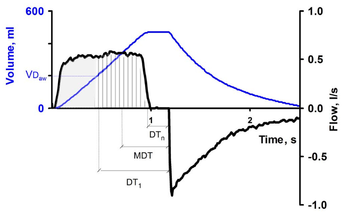
Also, these patients benefit from IRV, so don;t forget to use it. And increasing the end-inspiratory pause decreases the dead space ventilation, allowing lower tidal volumes for the same pCO2.
 
  • Like
Reactions: 1 user
Even without curves: if your PIP increases almost equally with your PEEP, even after 10-15 minutes, your PEEP is too high.

Do you mean driving pressure (plat minus PEEP) or do you mean PIP?

PIP (a measure of total pressures and an indicator of resistance) should increase (at least initially) with increasing PEEP. However, your driving pressure should decrease (ideally you are measuring transpulomonary driving pressure; but this is difficult with most vents available) and not increase as PEEP goes up (after a bit of time).

Please note, I am not lecturing here. This is my understanding and I am very open to being educated. Do you really mean PIP, FFP?

HH
 
  • Like
Reactions: 1 user
Even without curves: if your PIP increases almost equally with your PEEP, even after 10-15 minutes, your PEEP is too high.

Also, these patients benefit from IRV, so don;t forget to use it. And increasing the end-inspiratory pause decreases the dead space ventilation, allowing lower tidal volumes for the same pCO2.

I haven't manipulated the EIP before. If you prolong the EIP, is this added to your set I time (reducing your I:E) or is it a factor within the set I time (increasing your flow rate to achieve the same TV in a reduced time)? I'm imagining the former, since one study I found mentioned increasing PEEPi as a sequelae
 
Do you mean driving pressure (plat minus PEEP) or do you mean PIP?

PIP (a measure of total pressures and an indicator of resistance) should increase (at least initially) with increasing PEEP. However, your driving pressure should decrease (ideally you are measuring transpulomonary driving pressure; but this is difficult with most vents available) and not increase as PEEP goes up (after a bit of time).

Please note, I am not lecturing here. This is my understanding and I am very open to being educated. Do you really mean PIP, FFP?

HH
Please DO feel free to correct me. I am not a pulmonologist, hence ventilatory mechanics are definitely a weak point. But I think we are saying the same thing, just differently. I was using the poor anesthesiologist's method, who doesn't have Pplat or compliance measurement on the OR ventilator.

When one initially increases PEEP, one may also decrease dynamic compliance (hence increases PIP), especially in a stiff lung, unless the change in PEEP recruits new alveoli. In my OR experience, many times if I increase PEEP, the PIP will go up, too, at least for a short time. If some lung is recruited, or the lung and thoracic wall are compliant, the PIP will drop. If not, the PIP will stay high, many times with the same increase in PIP as the increase in PEEP.

I tried to find evidence of the above, but I couldn't, so please disregard.
 
I haven't manipulated the EIP before. If you prolong the EIP, is this added to your set I time (reducing your I:E) or is it a factor within the set I time (increasing your flow rate to achieve the same TV in a reduced time)? I'm imagining the former, since one study I found mentioned increasing PEEPi as a sequelae
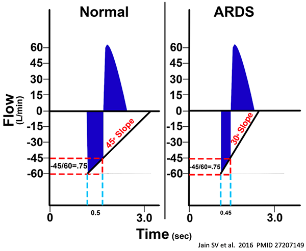
Unfortunately, I didn't bookmark the excellent explanation that was applying this concept to ARDS in Covid. It may have been on Twitter.

Regardless, here is a good article that explains the concept:
Key messages
  • In ARDS, CO2 exchange is importantly affected by the inspiratory flow wave pattern.
  • Mean distribution time (MDT) and end-inspiratory flow (EIF) influence of CO2 exchange, as expressed with a simple equation.
  • The effect of MDT is similar among ARDS patients, whereas that of EIF is variable.
  • A short insufflation followed by a long postinspiratory pause enhances CO2 exchange.
  • An efficient pattern of insufflation may be lung protective by allowing a lower tidal volume.

Found it, yay!
 
Last edited by a moderator:
  • Like
Reactions: 1 users
What makes you say that? (honest, not rhetorical question)

How do you determine "appropriate" PEEP? How do you determine "inappropriate" PEEP? Using FiO2? Using estimated or measured transpulmonary pressures? CXR? Oxygenation?

In my experience and observation, it is 10-100-fold more common to walk through an ICU and find insufficient PEEP than excess PEEP. Yes, the reason, as FFP has pointed out, people need PEEP 20 is often iatrogenic, but I don't think that is the point you are trying to make.

Although most docs are using more PEEP during the COVID hysteria (causing them to actually pay attention to PEEP and FiO2), I bet, despite this current environment, if you placed an esophageal balloon in every patient in the MICU and SICU tomorrow (assuming we are talking about adults in the US), you would not find more than one patient whose optimal PEEP was lower than the prescribed PEEP.

It is exceedingly rare (maybe a few times in my career) that I have found patients with such high PEEP that is over-distends to the point of vascular compromise, which is what I assume you are concerned about with a PEEP 20. In contrast, it is every day I am in the ICU that I find patients who have toxic FiO2, bibasilar atelectasis, unnecessary antibiotics, and tracheostomy for insufficient PEEP.

HH

There is a maximum o2 saturation that can be acheived by the patient..

The level of PEEP that gets you to that maximum Pa02 is your optimal peep

Lower than that number yields a lower Pao2 and thus is insufficient

Higher than that number and its too much and starts to shunt blood away

Practically, I adjust the peep in the OR until I reach the best O2sat that I can get

Im only doing this on patients with a low sat on 100% fio2.

Once I notice an improvement in sat with improved PEEP, I try to back off on the PEEP and see if I can maintain that sat

If they desaturate when I back off, I put the PEEP back up

Gradually, with little changes up and down, I try to hone in on that optimal peep setting
 
Last edited:
Members don't see this ad :)
Right, as in, if the paO2 doesnt require 100% fi02 , why are they prone at all? I would think that I would reserve turning prone (and all the complications that go with it) for those who have no other option, they are already maxed out on other oxygenation efforts..

can you provide evidence that it reduces mortality?

From what I have seen the evidence of reduced mortality is marginal even in a study with questionable methods

Here is an interesting one: Prone position for acute respiratory failure in adults. - PubMed - NCBI

And also, Im pretty sure that If i tried as hard as the guys who do most ICU studies, I could eek out a study that showed marginal benefit to recruitment manuevers and bronchodilators... or almost any intervention that i chose to study

I cant tell you how many times I have a patient with respiratory distress, no asthma history (but just had an LMA or a ETT in place) low sats, someone says "they are not wheezing" you give albuterol and they feel better and sats improve..

I think beta agonists are being underused from what Im seeing for that exact reasoning. You don't have to necessarily have a history of asthma or acute bronchospasm to get some oxygenation benefit from albuterol, especially when you have a viral infection of your airway causing some degree of reactivity.
Except people have tried numerous times to eek out a benefit of recruitment maneuvers and have failed. One can argue that the recruitment maneuver definition of 40/40 is too much, but there hasn't been a benefit shown. Which is more of a reflection of what you do in the OR not being translatable across an ICU population. A nuance few understand.
 
Please DO feel free to correct me. I am not a pulmonologist, hence ventilatory mechanics are definitely a weak point. But I think we are saying the same thing, just differently. I was using the poor anesthesiologist's method, who doesn't have Pplat or compliance measurement on the OR ventilator.

When one initially increases PEEP, one may also decrease dynamic compliance (hence increases PIP), especially in a stiff lung, unless the change in PEEP recruits new alveoli. In my OR experience, many times if I increase PEEP, the PIP will go up, too, at least for a short time. If some lung is recruited, or the lung and thoracic wall are compliant, the PIP will drop. If not, the PIP will stay high, many times with the same increase in PIP as the increase in PEEP.

I tried to find evidence of the above, but I couldn't, so please disregard.

Oh, I see where you are coming from...especially if you only have access to PIP measurements (and not plateaus). This will especially concern me if we end up using anesthesia machines during a surge. Hopefully we will be using them on patients without complicated pulmonary reasons for mechanical ventilation.

It is hard for me to explain without using plateau pressures. Let's use one of your scenarios to help me explain what I understand and experience. You said:

If not [alveoli not being recruited], the PIP will stay high, many times with the same increase in PIP as the increase in PEEP.

Yes, sometimes the PIP will increase exactly as much as the PEEP was raised. This new PIP (old PIP plus PEEP increase) often stays there (not much of a concern for me), but the driving pressure (plateau minus PEEP; which I care about a lot) will decrease. It is also possible that the driving pressure does not decrease. Both are possible and (without some other data, eg waveforms) it would be impossible to tell which. That is why plateaus are crucial -- for both management and comprehension of the (patho)physiology.

Again, this is just my understanding and experience. I am also not a pulmonologist (not that pulmonology fellowship is necessary or even helpful with the ventilator). FFP or others: please point out where I am mistaken. As you may be able to tell, I really like this stuff and find it helps patients tremendously. I want to be able to do this better, if I can learn something new.

HH
 
  • Like
Reactions: 1 users
Please DO feel free to correct me. I am not a pulmonologist, hence ventilatory mechanics are definitely a weak point. But I think we are saying the same thing, just differently. I was using the poor anesthesiologist's method, who doesn't have Pplat or compliance measurement on the OR ventilator.

When one initially increases PEEP, one may also decrease dynamic compliance (hence increases PIP), especially in a stiff lung, unless the change in PEEP recruits new alveoli. In my OR experience, many times if I increase PEEP, the PIP will go up, too, at least for a short time. If some lung is recruited, or the lung and thoracic wall are compliant, the PIP will drop. If not, the PIP will stay high, many times with the same increase in PIP as the increase in PEEP.

I tried to find evidence of the above, but I couldn't, so please disregard.
I disagree actually. There are three situations, with driving pressure being PIP/Plateau - PEEP:

1) Increasing PEEP leads to a decrease in PIP/Plateau pressure, this leads to an overall decrease in driving pressure and is indicative of recruitment of collapsed alveoli

2) Increasing PEEP leads to a commiserate increase in PIP/Plateau pressure, this leads to no change in driving pressure. In this situation you may not have recruited any alveoli, but you've optimized the PEEP in your open alveoli. No harm no foul.

3) Increasing PEEP leads to an increase in PIP/Plateau pressure that is greater than the magnitude by which your PEEP was increased. This leads to an increase in driving pressure. In this situation you have collapsed alveoli that were previously open and as such are causing barotrauma.

Ideally you want to be sitting at the tail end of scenario #2. If you're stuck in scenario 1 then you have not optimally recruited all recruitable alveoli.
 
  • Like
Reactions: 2 users
Except people have tried numerous times to eek out a benefit of recruitment maneuvers and have failed. One can argue that the recruitment maneuver definition of 40/40 is too much, but there hasn't been a benefit shown. Which is more of a reflection of what you do in the OR not being translatable across an ICU population. A nuance few understand.

My point is that you can prove anything if your study is determined to prove that thing..

There are actually studies on recruitment maneuvers claiming benefit..

Sometimes what Im doing in the OR "doesnt translate"

SOmetimes though, it does translate, and that is just an excuse to dismiss new ways of thinking/better ways to practice

Im not afraid to learn a thing or two from the ICU docs, but it goes both ways!
 
There is a maximum o2 saturation that can be acheived by the patient..

The level of PEEP that gets you to that maximum Pa02 is your optimal peep

Lower than that number yields a lower Pao2 and thus is insufficient

Higher than that number and its too much and starts to shunt blood away

Practically, I adjust the peep in the OR until I reach the best O2sat that I can get

Im only doing this on patients with a low sat on 100% fio2.

Once I notice an improvement in sat with improved PEEP, I try to back off on the PEEP and see if I can maintain that sat

If they desaturate when I back off, I put the PEEP back up

Gradually, with little changes up and down, I try to hone in on that optimal peep setting

Well, I am proposing that "titrating" PEEP to FiO2 or paO2 is not the way to do it. PEEP should be adjusted based on measured or estimated transplulmonary/transalveolar pressures.

Such adjustment has the beneficial "side effect" of allowing less FiO2 and better sat/paO2, but should not be driven by these outcomes...otherwise -- perhaps not during the short time in the OR (although detrimental effects are pretty quick) -- during an ICU stay, you end up with the problems I mentioned some of above: bibasilar atelectasis, toxic FiO2, unnecessary abx, higher driving pressures, prolonged mechanical ventilation, etc.

HH
 
  • Like
Reactions: 1 user
[...]

Again, this is just my understanding and experience. I am also not a pulmonologist (not that pulmonology fellowship is necessary or even helpful with the ventilator). FFP or others: please point out where I am mistaken. As you may be able to tell, I really like this stuff and find it helps patients tremendously. I want to be able to do this better, if I can learn something new.

HH
Me, too.

There are a ton of ways of optimizing PEEP. Deranged Physiology lists 10. I am sure many of us use most of the first 5, at least. (Using plateau pressures is #3).

  1. Use an arbitrarily high PEEP: set to 15-20cmH2O (CT studes support the use of a PEEP of around 16 cmH2O)
  2. Use the ARDSNet PEEP/FiO2 escalation tables (setting the PEEP according to the severity of the oxygenation failure)
  3. Titrate PEEP according to maximum compliance, i.e. set the PEEP which achieves the highest static compliance (this has the advantage of being tailored to each specific patient)
  4. Set the PEEP to slightly above the lower inflection point of the pressure volume curve (therefore avoiding cyclic atelectasis)
  5. Use a staircase recruitment (or derecruitment) manoeuvre to find the lowest PEEP at which the maximal oxygenation is maintained (this has the advantage of having a very pragmatic endpoint, SpO2)
  6. Titrate PEEP to achieve the smallest intrapulmonary shunt using a PA catheter with continuous SvO2 monitoring
  7. Titrate PEEP to achieve the lowest arterial minus end-tidal CO2 gradient (i.e. the PEEP at which dead space is minimal)
  8. Use the transpulmonary pressure calculated from oesophageal balloon manometry, using oesophageal pressure (Pes) as a surrogate for pleural pressure (where TPP = Pplat - Pes). Adjust PEEP so that TPP at end-expiration is 0-10.
  9. Using electrical impedance tomography, titrate PEEP to achieve the highest electrical impedance in the thorax (i.e. the greatest amount of aerated lung)
  10. Use sequential CT scans to visually determine a PEEP at which the greatest volume of lung is recruited during end-expiration"
 
I disagree actually. There are three situations, with driving pressure being PIP/Plateau - PEEP:

1) Increasing PEEP leads to a decrease in PIP/Plateau pressure, this leads to an overall decrease in driving pressure and is indicative of recruitment of collapsed alveoli

2) Increasing PEEP leads to a commiserate increase in PIP/Plateau pressure, this leads to no change in driving pressure. In this situation you may not have recruited any alveoli, but you've optimized the PEEP in your open alveoli. No harm no foul.

3) Increasing PEEP leads to an increase in PIP/Plateau pressure that is greater than the magnitude by which your PEEP was increased. This leads to an increase in driving pressure. In this situation you have collapsed alveoli that were previously open and as such are causing barotrauma.

Ideally you want to be sitting at the tail end of scenario #2. If you're stuck in scenario 1 then you have not optimally recruited all recruitable alveoli.
Actually, upon reviewing my notes, I'm mistaken. The scenarios are:

1) Driving pressure decreases
2) Driving pressure increases, but less than the magnitude by which you increased your PEEP.
3) Driving pressure increases commiserate with your PEEP and that means you haven't recruited anything.

Which might have been what you were saying FFP. Keep going up on PEEP until plateau/PIP increases in a 1:1 fashion.

Sorry.
Yep, except that PIP is a dynamic measure, hence it also involves airway resistance, while plateau is static, so it's more "scientific".
 
Last edited by a moderator:
  • Like
Reactions: 1 user
There is a maximum o2 saturation that can be acheived by the patient..

The level of PEEP that gets you to that maximum Pa02 is your optimal peep

Lower than that number yields a lower Pao2 and thus is insufficient

Higher than that number and its too much and starts to shunt blood away

Practically, I adjust the peep in the OR until I reach the best O2sat that I can get

Im only doing this on patients with a low sat on 100% fio2.

Once I notice an improvement in sat with improved PEEP, I try to back off on the PEEP and see if I can maintain that sat

If they desaturate when I back off, I put the PEEP back up

Gradually, with little changes up and down, I try to hone in on that optimal peep setting

Since you're talking about PEEP in the OR, where do you stop with SLV?
 
Me, too.

There are a ton of ways of optimizing PEEP. Deranged Physiology lists 10. I am sure many of us use most of the first 5, at least. (Using plateau pressures is #3).

  1. Use an arbitrarily high PEEP: set to 15-20cmH2O (CT studes support the use of a PEEP of around 16 cmH2O)
  2. Use the ARDSNet PEEP/FiO2 escalation tables (setting the PEEP according to the severity of the oxygenation failure)
  3. Titrate PEEP according to maximum compliance, i.e. set the PEEP which achieves the highest static compliance (this has the advantage of being tailored to each specific patient)
  4. Set the PEEP to slightly above the lower inflection point of the pressure volume curve (therefore avoiding cyclic atelectasis)
  5. Use a staircase recruitment (or derecruitment) manoeuvre to find the lowest PEEP at which the maximal oxygenation is maintained (this has the advantage of having a very pragmatic endpoint, SpO2)
  6. Titrate PEEP to achieve the smallest intrapulmonary shunt using a PA catheter with continuous SvO2 monitoring
  7. Titrate PEEP to achieve the lowest arterial minus end-tidal CO2 gradient (i.e. the PEEP at which dead space is minimal)
  8. Use the transpulmonary pressure calculated from oesophageal balloon manometry, using oesophageal pressure (Pes) as a surrogate for pleural pressure (where TPP = Pplat - Pes). Adjust PEEP so that TPP at end-expiration is 0-10.
  9. Using electrical impedance tomography, titrate PEEP to achieve the highest electrical impedance in the thorax (i.e. the greatest amount of aerated lung)
  10. Use sequential CT scans to visually determine a PEEP at which the greatest volume of lung is recruited during end-expiration"

There are a couple minor methodological issues with the ART trial, but based on its signal toward increased mortality, #5 staircase recruitment has really fallen out of favor. I think manual recruitment maneuvers probably have some value in ARDS, but I would definitely avoid some of the "start at crazy high PEEP and titrate down" protocols.

 
You say determining optimum peep is not hard science. How do you do it? I just use a PEEP table cuz it’s easier, but if you have a better way, I’d love to learn

I put into a pressure control mode and keep the same driving pressure while changing PEEP and find the point where the compliance is the greatest. I take into consideration the obesity of the patient and the hemodynamic effect too-low cardiac output can worsen oxygenation/ventilation through low SVO2/increased dead space respectively, and more compliant lungs transmit more pressure to the veins.

Ideally done while paralysed so that you eliminate the patient effort component of trans pulmonary pressure (since we don’t use oesophageal manometers)

 
  • Like
Reactions: 1 user
One can also get an idea of compliance by titrating based on stress index

DP-3.png


Yep, except that PIP is a dynamic measure, hence it also involves airway resistance, while plateau is static, so it's more "scientific".
Yeah, I just use the two interchangeably depending on if you're in a PC or VC mode of ventilation.

The big problem with this is the time intense nature of the calibration. You need some sort of recruitment maneuver followed by a PEEP adjustment, then reassessing a little while later. And continuing to do that until you find the right PEEP.

Which might be why I'm starting to lean towards APRV as a primary mode of ventilation in ARDS. The problem with APRV is most people haphazardly choose the parameters associated with it, rather than using a physiologically based approach.
 
Since you're talking about PEEP in the OR, where do you stop with SLV?

I essentially do the same thing with SLV, but when I cant get a decent sat despite every PEEP setting I try and 100% Fio2, I use CPAP to the down (surgical) lung. I give as much CPAP to the down lung as I can, only limited by the lung blowing up in the surgeons field and reducing visualization
 
Yeah, I just use the two interchangeably depending on if you're in a PC or VC mode of ventilation.

The big problem with this is the time intense nature of the calibration. You need some sort of recruitment maneuver followed by a PEEP adjustment, then reassessing a little while later. And continuing to do that until you find the right PEEP.

Which might be why I'm starting to lean towards APRV as a primary mode of ventilation in ARDS. The problem with APRV is most people haphazardly choose the parameters associated with it, rather than using a physiologically based approach.

whats your approach to aprv?
 
whats your approach to aprv?

The key is setting the T low to 75% of expiratory flow to prevent derecruitment.
 
  • Like
Reactions: 1 users
This is a great discussion. Thanks to all who are participating. After spending some time in SPF, this is refreshing.

There is a lot to respond to here; and I hope to get a chance soon, but clinical responsibilities may get in the way.

...but I have time at least to propose a question to the APRV fan club:

Why is a Phigh of 30 not concerning but a PEEP 25 so deeply concerning regarding venous return and alveolar capillary compression?

HH

I wouldn't say a Th of 30 is unconcerning but Tl is an opportunity for venous return not there on conventional vent mode
 
This is a great discussion. Thanks to all who are participating. After spending some time in SPF, this is refreshing.

There is a lot to respond to here; and I hope to get a chance soon, but clinical responsibilities may get in the way.

...but I have time at least to propose a question to the APRV fan club:

Why is a Phigh of 30 not concerning but a PEEP 25 so deeply concerning regarding venous return and alveolar capillary compression?

HH
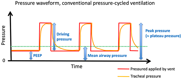
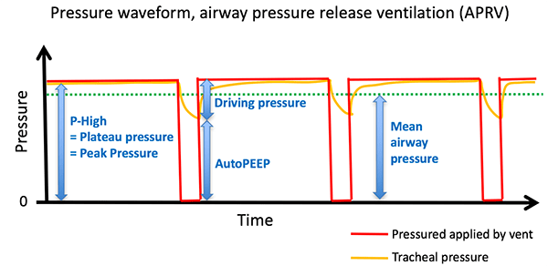
I believe it has to do with mean airway pressure more so than one isolated number. In APRV, with a pHigh of 30, your mean airway pressure will never be more than 30 (It'll actually be less based off the number of releases per minute). If you have a PEEP of 25, and a driving pressure of ~15, then your mean airway pressure is going to be hanging out a lot closer to 40.

I will also say that the concerns about PEEP and venous return are probably overblown. I'm sure someone else can augment this explanation though.
 
  • Like
Reactions: 1 users
I believe it has to do with mean airway pressure more so than one isolated number. In APRV, with a pHigh of 30, your mean airway pressure will never be more than 30 (It'll actually be less based off the number of releases per minute). If you have a PEEP of 25, and a driving pressure of ~15, then your mean airway pressure is going to be hanging out a lot closer to 40.

I will also say that the concerns about PEEP and venous return are probably overblown. I'm sure someone else can augment this explanation though.

gr1aprv.png


https://i2.wp.com/emcrit.org/wp-content/uploads/2017/11/gr2aprv.png


Source: PulmCrit- APRV: Resurrection of the open-lung strategy?
 
Last edited by a moderator:
Correct me if I’m wrong, if patients are on APRV and breathing spontaneously at Phigh, if they have on PSV or ATC, aren’t they generating pressures above Phigh?
 
Correct me if I’m wrong, if patients are on APRV and breathing spontaneously at Phigh, if they have on PSV or ATC, aren’t they generating pressures above Phigh?
You're right. I am not an APRV user, but I remember that PSV and ATC are not recommended during APRV, exactly because of the concerns regarding transpulmonary pressure and VILI. Plus it's debatable whether PSV doesn't defeat the benefits of SV during APRV.
 
Last edited by a moderator:
Correct me if I’m wrong, if patients are on APRV and breathing spontaneously at Phigh, if they have on PSV or ATC, aren’t they generating pressures above Phigh?

APRV can be well tolerated for a spontaneously breathing pt but a significant amount of PSV should not be used


7BAC827A-65D3-44DE-A99F-C8DD9EB0CA70.jpeg
 
  • Like
Reactions: 2 users
What makes you say that? (honest, not rhetorical question)

How do you determine "appropriate" PEEP? How do you determine "inappropriate" PEEP? Using FiO2? Using estimated or measured transpulmonary pressures? CXR? Oxygenation?

In my experience and observation, it is 10-100-fold more common to walk through an ICU and find insufficient PEEP than excess PEEP. Yes, the reason, as FFP has pointed out, people need PEEP 20 is often iatrogenic, but I don't think that is the point you are trying to make.

Although most docs are using more PEEP during the COVID hysteria (causing them to actually pay attention to PEEP and FiO2), I bet, despite this current environment, if you placed an esophageal balloon in every patient in the MICU and SICU tomorrow (assuming we are talking about adults in the US), you would not find more than one patient whose optimal PEEP was lower than the prescribed PEEP.

It is exceedingly rare (maybe a few times in my career) that I have found patients with such high PEEP that is over-distends to the point of vascular compromise, which is what I assume you are concerned about with a PEEP 20. In contrast, it is every day I am in the ICU that I find patients who have toxic FiO2, bibasilar atelectasis, unnecessary antibiotics, and tracheostomy for insufficient PEEP.

HH
So how do you define optimal peep then?

As someone who spent an entire summer of fellowship shoving esophageal manometers into patients, plus lung USS, diaphragm uss impedance probes im interested
 
I find all of these discussions re ventilation strategies interesting but i have yet to see a single mode to have any statistically significant benefit except ardsnet and even that had plenty of flaws. PAV, APRV, NAVA any of them have any reasonable evidence that one is better than the other?
 
  • Like
Reactions: 1 user
I find all of these discussions re ventilation strategies interesting but i have yet to see a single mode to have any statistically significant benefit except ardsnet and even that had plenty of flaws. PAV, APRV, NAVA any of them have any reasonable evidence that one is better than the other?

 
Does anyone else look at trials coming out of China and think “I’ll just wait for an American study”? Or am I the only one who has little faith in their research...
 
  • Like
Reactions: 2 users
To the OP - an unsolvable problem that you shouldn't try to solve.

Morbidly obese, ARDS, proning, can't/won't trach, mortality north of 80-90%.

Primum non nocere. The patient is the one with the disease.

Why won't ENT trach? Bedside trach? [all bad options]
 
Does anyone else look at trials coming out of China and think “I’ll just wait for an American study”? Or am I the only one who has little faith in their research...

That study was pretty well designed, but regardless of whatever China skepticism there is I don't think anyone should take a single center study from anywhere as gospel. I'm just pointing out that there is some evidence that APRV works.
 
2 vs 19 ventilator free days? Colour me skeptical. 14 percent mortality difference? We’ve never found such a (real) mortality difference for anything in the ICU.

APRV is great if you know how to use it. It’s all institution dependent though. My institution is not familiar, so we would probably kill people if we put them on APRV. Other institutions that use APRV may not know how to use VC properly. It’s more dependent on nursing staff (or RTs I guess for you guys) who are really the ones responsible for ensuring adherence to a ventilation strategy, because we can’t be at the beside 24/7. If they don’t understand what’s being done, the whole ventilation plan is doomed to fail.
 
  • Love
Reactions: 1 user
2 vs 19 ventilator free days? Colour me skeptical. 14 percent mortality difference? We’ve never found such a (real) mortality difference for anything in the ICU.

APRV is great if you know how to use it. It’s all institution dependent though. My institution is not familiar, so we would probably kill people if we put them on APRV. Other institutions that use APRV may not know how to use VC properly. It’s more dependent on nursing staff (or RTs I guess for you guys) who are really the ones responsible for ensuring adherence to a ventilation strategy, because we can’t be at the beside 24/7. If they don’t understand what’s being done, the whole ventilation plan is doomed to fail.
xigris made a 30% mortality difference in sepsis didn't it :whistle:
 
2 vs 19 ventilator free days? Colour me skeptical. 14 percent mortality difference? We’ve never found such a (real) mortality difference for anything in the ICU.

APRV is great if you know how to use it. It’s all institution dependent though. My institution is not familiar, so we would probably kill people if we put them on APRV. Other institutions that use APRV may not know how to use VC properly. It’s more dependent on nursing staff (or RTs I guess for you guys) who are really the ones responsible for ensuring adherence to a ventilation strategy, because we can’t be at the beside 24/7. If they don’t understand what’s being done, the whole ventilation plan is doomed to fail.

Yes, you should be skeptical. Single-center studies are jumping off points to see if anyone can actually replicate the findings.

Here is a good critique of the Zhou study


APRV for ARDS: the complexities of a mode and how it affects even the best trials
 
  • Like
Reactions: 1 users


The whole thread is worth reading:


This actually happened in the other room while I was emergently intubating another room.

Makes me wonder:
Has anyone diagnosed this in real life based solely on vent parameters? Or has anyone's suspicions from vent parameters alone caused anyone to investigate the tube?

Is anyone checking daily peak and plateau pressures and paying attention time constant (or RC?).

For his example here:
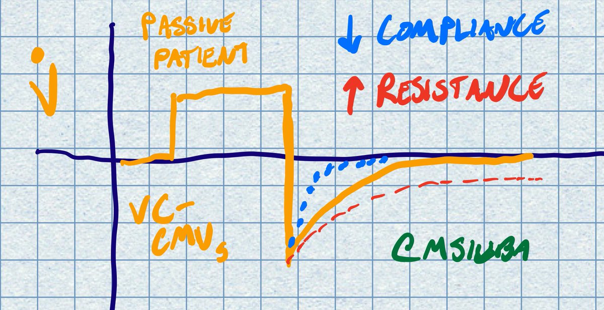
EVIZTzeXgAUwtIj.jpg


How do you actually notice the difference between the red and the yellow line in clinical practice?


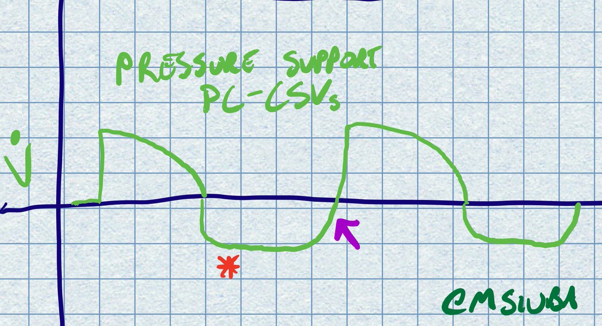
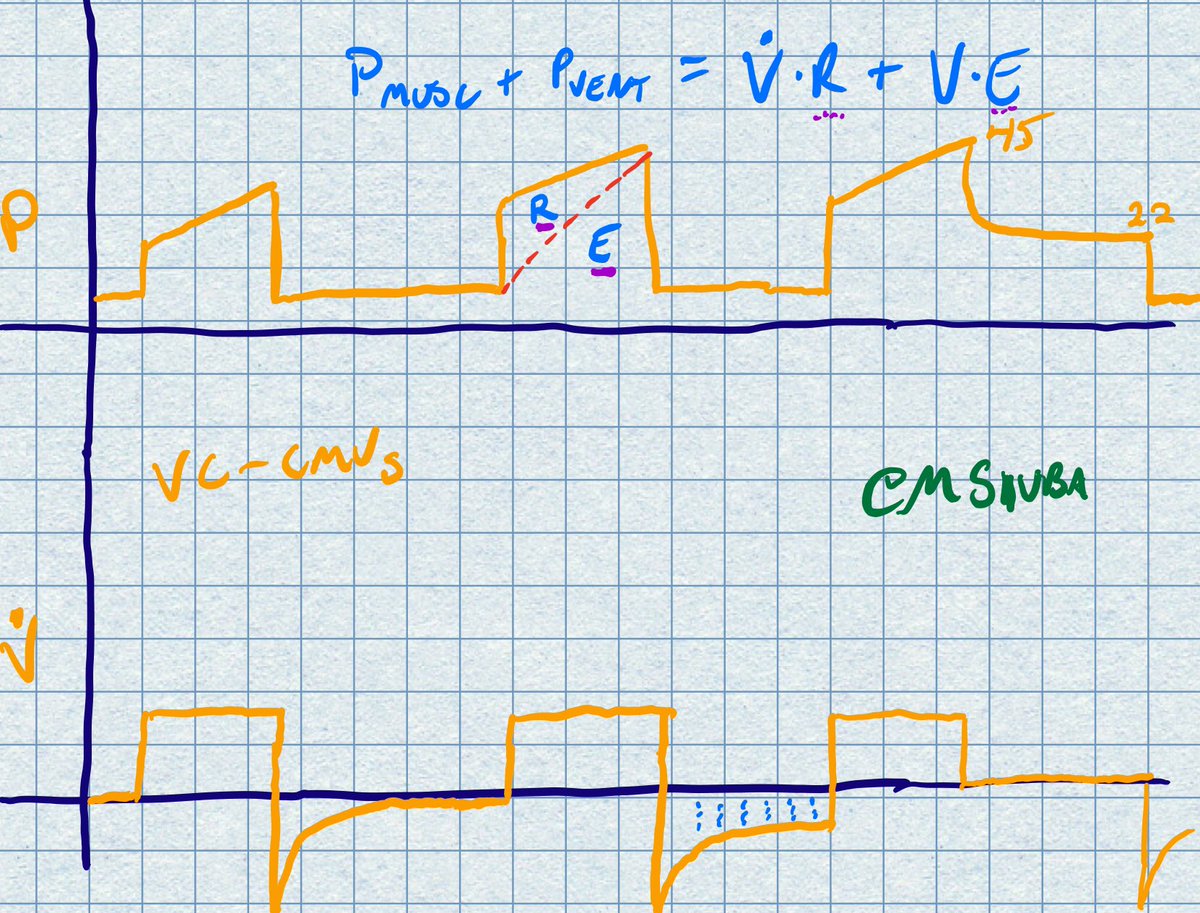
These two images also tell a thousand words, i'm gonna watch out for this in the future:

EVIaItGXYAATbic.jpg


EVIa2DUXYAIdaRV.jpg


Lastly, we were just talking about how Pressure control mode with a constant pressure and variable flow is best for the pathophysiology, but all these diagnostics are in volume control mode. Is there a way to diagnose this in a constant pressure mode? (can you insp pause on pressure control mode?)
 
Last edited:
This actually happened in the other room while I was emergently intubating another room.

Makes me wonder:
Has anyone diagnosed this in real life based solely on vent parameters? Or has anyone's suspicions from vent parameters alone caused anyone to investigate the tube?

Is anyone checking daily peak and plateau pressures and paying attention time constant (or RC?).

For his example here:
EVIZTzeXgAUwtIj.jpg


How do you actually notice the difference between the red and the yellow line in clinical practice?


These two images also tell a thousand words, i'm gonna watch out for this in the future:

EVIaItGXYAATbic.jpg


EVIa2DUXYAIdaRV.jpg


Lastly, we were just talking about how Pressure control mode with a constant pressure and variable flow is best for the pathophysiology, but all these diagnostics are in volume control mode. Is there a way to diagnose this in a constant pressure mode? (can you insp pause on pressure control mode?)


One could easily diagnose and treat it with a bronchoscope but I imagine that’s just not happening with COVID patients.
 
  • Sad
Reactions: 1 user
This actually happened in the other room while I was emergently intubating another room.

Makes me wonder:
Has anyone diagnosed this in real life based solely on vent parameters? Or has anyone's suspicions from vent parameters alone caused anyone to investigate the tube?

Is anyone checking daily peak and plateau pressures and paying attention time constant (or RC?).

For his example here:
EVIZTzeXgAUwtIj.jpg


How do you actually notice the difference between the red and the yellow line in clinical practice?


These two images also tell a thousand words, i'm gonna watch out for this in the future:

EVIaItGXYAATbic.jpg


EVIa2DUXYAIdaRV.jpg


Lastly, we were just talking about how Pressure control mode with a constant pressure and variable flow is best for the pathophysiology, but all these diagnostics are in volume control mode. Is there a way to diagnose this in a constant pressure mode? (can you insp pause on pressure control mode?)

Easy enough to switch quickly into VC, see if there’s a large difference between pip and plateau, then switch back to PC.

You can inspiration pause on PC by making the i time really long (or doing inspiration hold maneuover)- eventually your flow will become zero and then pip becomes equal to plateau. But the whole PIP/plateau concept is invalid in pressure control because of the decelerating flow.
 
  • Like
Reactions: 1 user
Top