Help interpreting dental radiographs

This forum made possible through the generous support of SDN members, donors, and sponsors. Thank you.
Status
Not open for further replies.

Dnaia

New Member
5+ Year Member
Joined
Nov 25, 2017
Messages
4
Reaction score
2
Hi everyone...
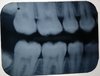
I have an oral test interpreting these two radiographs tomorrow, and I was wondering if you could help, in case I missed anything.

What the structure above the maxillary sinus in the periapical? My first thought would be the zygomatic process, but shouldn't that be a bit lower?

Thanks in advance,
Dania

IMG_20171125_200806.jpg
IMG_20171125_200932.jpg

Members don't see this ad.
 
  • Like
Reactions: 1 user
Hi Dania

I think you are correct !
that is the zygomatic process

good Luck with your Exam
 
  • Like
Reactions: 1 users
Hi Dania

I think you are correct !
that is the zygomatic process

good Luck with your Exam

Yay thanks! Did you notice anything else of significance that I may be asked about?

Thank you again
 
  • Like
Reactions: 1 user
Members don't see this ad :)
Yay thanks! Did you notice anything else of significance that I may be asked about?

Thank you again

I recommend you to focus on the other anatomical and radiographical landmarks. Ex:periodontal space , lamina dura and pulp space .

Regarding the periapical radiograph
There is proximal caries i.r.t #3

Regarding the bite-wing one :
There is proximal caries i.r.t the lower 1st premolar

clearly there is also impaction of the 3rd molars
 
  • Like
Reactions: 1 user
I recommend you to focus on the other anatomical and radiographical landmarks. Ex:periodontal space , lamina dura and pulp space .

Regarding the periapical radiograph
There is proximal caries i.r.t #3

Regarding the bite-wing one :
There is proximal caries i.r.t the lower 1st premolar

clearly there is also impaction of the 3rd molars
Thank you so much! I really appreciate it! My professor saw very superficial caries on the proximal surfaces of the upper premolars on the bitewing, but I couldn't see anything, can you?

Also, could you look at these two radiographs of it's not an inconvenience?
IMG_20171125_232817.jpg
IMG_20171125_232907.jpg
 

Attachments

  • IMG_20171125_200932.jpg
    IMG_20171125_200932.jpg
    70.4 KB · Views: 44
I am Happy to help !

The way to detect the proximal caries radiographically is to observe any radiolucencies in the dentin and enamel in the proximal region .

Regarding the first radiograph there is incisal tooth loss i.r.t #31 and #41. There is also vertical defect proximally between the two incisors.It might be periodontal Abscess , but it needs more investigation.

With regards to the second one . There is widen periodontal space i.r.t the mesial side of the lower second molar . There is also proximal caries between the upper and lower premolars .
 
SDN is not for medical advice or HW help. Closing.
 
  • Like
Reactions: 1 user
Status
Not open for further replies.
Top