- Joined
- Dec 29, 2012
- Messages
- 247
- Reaction score
- 4
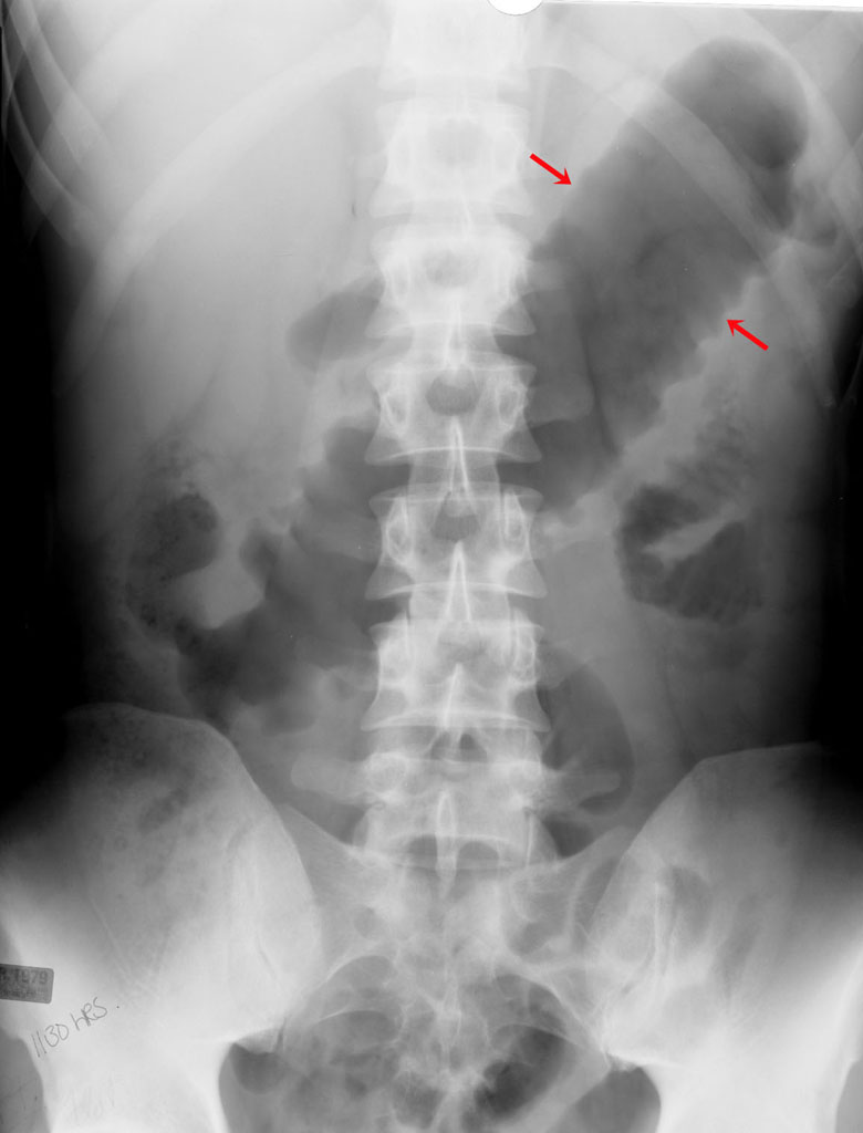
68 yo male patient presents to your clinic with postprandial pain. X-ray below, whats the dx?

aortic coarcatation
I have no clue. tough one for sure
horseshoe kidney 😳
you knew it...esp when given answer choices
 That's crazy though. Good one!
wayyyyyyyy off
7 days old dyspnea/ hypoplastic lungs due to diaphgramatic hernia.

1) What could have caused this?
2) What's the SaO2 in this pt (i.e. increased/decreased?)
crazy, eh...always used to seeing in in x-ray point of view...here's my lunch time contributions
dx
looking for the complication, of this dx
bonus points if you can give me the disease it's found in
Toxic Megacolon
Complications could be perforation (leading to peritonitis), sepsis, dehydration from chronic diarrhea.
Related to C. Diff colitis with pseudomembranes.
Also, I once had a patient with C. Diff Ileitits (which I didn't even know was possible).
7 days old dyspnea/ hypoplastic lungs due to diaphgramatic hernia.
1) What could have caused this?
2) What's the SaO2 in this pt (i.e. increased/decreased?)
👍
didnt know CDif could cause this, but that's cool. was looking for inflammatory bowel disease...specifically, ulcerative...😳

Hashimoto's - looks like appearance of a germinal center...& possible scalloping on the right side
Hashimoto's - looks like appearance of a germinal center...& possible scalloping on the right side
thanks!
this actually helps me understand wikipedias description
" retrograde (reversed) flow of blood in the vertebral artery or the internal thoracic artery, due to a proximal stenosis (narrowing) and/or occlusion of the subclavian artery. The arm may be supplied by blood flowing in a retrograde direction down the vertebral artery at the expense of the vertebrobasilar circulation. This is called the subclavian steal."
Figured its about time for me to join in on this fun instead of just lurking.
It does look like a germinal center, but I don't think that's scalloping. That would be with Grave's disease anyway, no scalloping in Hashimoto's I believe?

Does that look like a granuloma to anyone? I was thinking granulomatous thyroiditis.
Does that look like a granuloma to anyone? I was thinking granulomatous thyroiditis.
Yeah, thats what I was thinking too haha.

macrophagepoint to it...because i don't see it...would be helpful to the others...if not
here's my question:
what cell is responsible for the changes seen in the lung...
hint: not looking for what the picture below is...
point to it...because i don't see it...would be helpful to the others...if not
here's my question:
what cell is responsible for the changes seen in the lung...
hint: not looking for what the picture below is...
macrophage
Fibroblasts, which are stimulated by the macrophages which ingest the asbestos fibers.
Causes interstitial pulmonary fibrosis, and there's a risk of mesothelioma and other lung CA as well.
nice fellas...
i think theres a typo in pathoma...he talks about T2Pneumocytes are responsible for causing fibrosis...
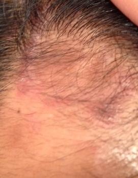
btw, who here knows their skin pathology really really well...to the point they're banking on getting skin questions because they feel confident they'll get it right...
I know melenoma, bcc, scc, psoriasis, and urticaria...what else is there to know?? 😎
point to it...because i don't see it...would be helpful to the others...if not

I don't know what in particular lol it's just that the supposed follicle looked like a granuloma. It even looks like there could be a giant cell in there.
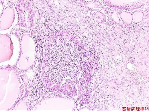
(I had to cheat and look this one up to be sure: http://oac.med.jhmi.edu/pathconcepts/ShowImage.cfm?TutorialID=5&ConceptID=21&ImageID=136 )
No granuloma according to that, it is Hashimoto's. On the right is the follicle, with the germinal center that was already pointed out on the left.
yeah i guess at our level that's about it...and the blisters...was hoping somebody here has a good thorough skin knowledge...wanted to ask a personal question actually
well post it... for educational purposes
Post the Q mate

yeah i guess at our level that's about it...and the blisters...was hoping somebody here has a good thorough skin knowledge...wanted to ask a personal question actually
It's herpes, Jonari
too shy...it's ok, thanks anyways though fellas 😳
ok lets get back on topic

whats dis?
ok lets get back on topic
whats dis?
ok lets get back on topic
whats dis?
Looks like scabies to me
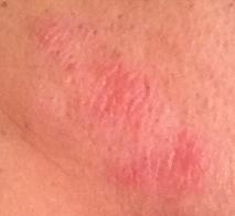
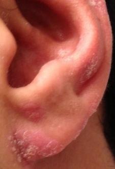
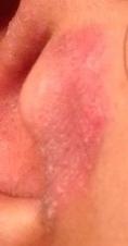
I want to say Giardia? Although it doesn't exactly match what I'm picturing since this is a little more round.
seborrheic dermatitis? I get something similar around my nose, especially when im stressed out. 2% hydrocortisone clears it right up
cot dam it....i was hoping it wasn't that...fml
lol take my guess with a grain of salt... it just seems similar to what i get. although I mostly get it around my nose. A little hydrocortisone once or twice a week when its really bad clears it up in no time.