
-
CycleTrack and SDN are teaming up to make medical school admissions more transparent and accessible! Read the announcement to learn how we’re supporting the future of CycleTrack’s free application-tracking tools .
You are using an out of date browser. It may not display this or other websites correctly.
You should upgrade or use an alternative browser.
You should upgrade or use an alternative browser.
Step I USMLE images
Started by Zuhal

Get help with your application
Use all the free resources available to you from SDN: articles, guides, expert advising, forums discussions, and school research.


yeah...no, i came to the same conclusion and the derm said the same thing too...hopefully it'll go away when the test is done...thanks for the insight...back on topic now 😳
Sure it's the water and not your soap? Also try a water softner, you could be allergic to one of those heavy metals and others aren't.
Advertisement - Members don't see this ad
Sure it's the water and not your soap? Also try a water softner, you could be allergic to one of those heavy metals and others aren't.
will do thanks...😀👍
psoriasis...able to pull the nail off easily
nope... psoriasis is more associated with pitting and other really freaky looking features

ok to keep with the derm theme

whats this called? and what can it be associated with?
Zinc deficiency? I know that causes nail changes, not sure what they look like though.

ok to keep with the derm theme
whats this called? and what can it be associated with?
Mee's lines, arsenic poisoning?
There's a Beau's line near the tip of the lunula too.
Edit, maybe they're both Beau's, I haven't looked at a lot of these.
Zinc deficiency? I know that causes nail changes, not sure what they look like though.
no... this is a line that has a particular name
Mee's lines, arsenic poisoning?
There's a Beau's line near the tip of the lunula too.
Its Beau's line, not mees though... they look different.
Systemic disease implicated if all 20 nails involved. Pathogenesis: Occur after any severe, sudden, acute, particularly febrile illness; damage to matrix. Etiology: High fever, postnatal, cytotoxic drugs, severe adverse cutaneous drug reaction. Findings: Transverse, bandlike depressions in nail, extending from one lateral edge to the other, affecting all nails at corresponding levels (Fig. 33-23). If duration of disease completely inhibits matrix activity for 714 days, transverse depression results in total division of nail plate (onychomadesis). Multiple parallel lines with chemotherapy. Duration: Thumbnails (lines present for 69 months) and large nails (lines present for up to 2 years) are most reliable markers.
from clinical derm atlas
Its Beau's line, not mees though... they look different.
from clinical derm atlas
Yeah, I've only seen them look like a clear depression (like the one nearest the cuticle) never seen one that was pale colored like the one in the mid-nail. It's tough to see if there's a depression there or not.
Mee's lines are pale bands through the nail, a lot like that image, but everyone I've seen had faint borders.
But the pale one could (and probably does) represent a more severe attack of whatever this person has that's causing the lines, it looks like the nail almost split.

1. paraesophageal hernia
2. SaO2 = saturation of rbcs with hemoglobin, right? other lung is still working fine and being able to oxygenate the rbcs...so shouldn't it be normal?
1) Defective development of the pleuroperitoneal membrane (which becomes the diaphragm)
2) I gotta think the SaO2 is decreased.
No. Does anyone wanna give it a try before I tell you the answer?
No. Does anyone wanna give it a try before I tell you the answer?
Is it a Bochdalek hernia?
Last edited:
No. Does anyone wanna give it a try before I tell you the answer?
is this something about how the organs herniate in the posterlateral style into the retroperitoneum by any chance? i remember reading something similar to this in goljan's rr
wait what are you talking about?
post #754 on the previous page
Advertisement - Members don't see this ad
is this something about how the organs herniate in the posterlateral style into the retroperitoneum by any chance? i remember reading something similar to this in goljan's rr
Yea thats called a Bochdalek hernia, I dont know if thats what Zuhal is looking for?

34 year old presents to E.R. with fever, jaundice, and anorexia. Has upper right quad pain.
here is the liver biopsy: What is this type of cell, what does it contain, and what is the disease?

edit: sorry picture is so big. black thing is an arrow pointing to the pathology.
here is the liver biopsy: What is this type of cell, what does it contain, and what is the disease?
edit: sorry picture is so big. black thing is an arrow pointing to the pathology.
ok, one more before I peace out for the night:
Patient has recurrent renal colic involving general abdomen pain and recently has had some renal stones.
xray of head, what is the dx?

Patient has recurrent renal colic involving general abdomen pain and recently has had some renal stones.
xray of head, what is the dx?
ok, one more before I peace out for the night:
Patient has recurrent renal colic involving general abdomen pain and recently has had some renal stones.
xray of head, what is the dx?
Paget's?
Paget's?
Nope, but good guess.
ok, one more before I peace out for the night:
Patient has recurrent renal colic involving general abdomen pain and recently has had some renal stones.
xray of head, what is the dx?
Multiple myeloma?
Nope, but good guess.
Hyperparathyroidism?
I forgot Paget's presents with normocalcemia since it's a remodeling problem, not a pure osteolysis problem.

ok, one more before I peace out for the night:
Patient has recurrent renal colic involving general abdomen pain and recently has had some renal stones.
xray of head, what is the dx?
Punched out lytic lesions..multiple myeloma
34 year old presents to E.R. with fever, jaundice, and anorexia. Has upper right quad pain.
here is the liver biopsy: What is this type of cell, what does it contain, and what is the disease?
edit: sorry picture is so big. black thing is an arrow pointing to the pathology.
Kupffer cells, in alcoholic hepatitis?
Hyperparathyroidism?
I forgot Paget's presents with normocalcemia since it's a remodeling problem, not a pure osteolysis problem.
that's it, great work.
This is salt and pepper calavarium, which is periosteal thinning.
I am actually not sure how to differentiate this form multiple myeloma.... both would presents with hypercalcemia and bone structural changes...hm. I guess it requires looking for an M spike.
Kupffer cells, in alcoholic hepatitis?
no but close, this is a kupffer cell:

that's it, great work.
This is salt and pepper calavarium, which is periosteal thinning.
I am actually not sure how to differentiate this form multiple myeloma.... both would presents with hypercalcemia and bone structural changes...hm. I guess it requires looking for an M spike.
i know most don't do kaplan Q bank but when i did it...there was a question that had both answer choices up there...it also included that the ESR was elevated, talked about doing a biopsy showed congo red stain, and in the urine you saw features "which were indicative of features found on an antibody structure".
so i guess seeing those extra clues are needed to differentiate between the two
34 year old presents to E.R. with fever, jaundice, and anorexia. Has upper right quad pain.
here is the liver biopsy: What is this type of cell, what does it contain, and what is the disease?
edit: sorry picture is so big. black thing is an arrow pointing to the pathology.
Ito cell aka ? (brain fart atm would get it out of a multiple choice answer though) Contains Vitamin A.
Ito cell aka ? (brain fart atm would get it out of a multiple choice answer though) Contains Vitamin A.
I'll give it up, I think it's a rather obscure pathology but I got a UW question on it.
It's a councilman body, it has acidophilic granules and indicates apoptosis. They are seen are a viral hepatic infection (most likely hep B or C in this case due to drug use). I think they go along with spaces int he liver and hepatocyte balloning.
http://en.wikipedia.org/wiki/Councilman_body
another pic:

Advertisement - Members don't see this ad
I'll give it up, I think it's a rather obscure pathology but I got a UW question on it.
It's a councilman body, it has acidophilic granules and indicates apoptosis. They are seen are a viral hepatic infection (most likely hep B or C in this case due to drug use). I think they go along with spaces int he liver and hepatocyte balloning.
http://en.wikipedia.org/wiki/Councilman_body
another pic:
I would think these are actually pretty high yield - they show most of the typical features of apoptosis
arrow is pointing to what? - ive heard 2 different answers, so hopefully somebody can clear it up.
dx?
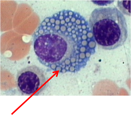
Those look like the cytoplasmic Ig inclusions of Multiple Myeloma to me.
Those look like the cytoplasmic Ig inclusions of Multiple Myeloma to me.

any idea what they're called...ok so it's Ig inclusions, right? i've heard somebody else saying that their ER accumulations...so was confused
any idea what they're called...ok so it's Ig inclusions, right? i've heard somebody else saying that their ER accumulations...so was confused
Accumulation of RER product maybe. +1 for the Kip gif
Accumulation of RER product maybe. +1 for the Kip gif
yeah that's probably what they were trying to convey...thanks!
are those neurofibrillary tangles? Intracellular hyperphosphorylated Tau protein, with alzheimer's, chromosome 21. Don't recall how to slow it down though...dx?
chromosomes involved?
treatments to slow down?
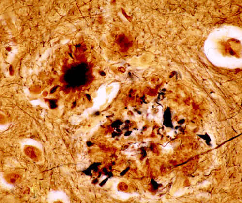
are those neurofibrillary tangles? Intracellular hyperphosphorylated Tau protein, with alzheimer's, chromosome 21. Don't recall how to slow it down though...
👍, right dx of alzheimer's...but that's Abeta amyloid...didnt know what it was because i've always seen questions with this type of picture
http://upload.wikimedia.org/wikiped...on_with_Alzheimer-related_pathology,_HE_3.JPG
yup, chromosome 21 causes the Abeta deposition...and other chromosomes for alzheimer's include: 1 and 14 for early onset, and 19 for late onset.
you can help alzheimer's pts by giving achesterase inhibitor (ie donepezil, tacarine) and can also give galantamine (sp?)...doesnt do anything with respect to amyloid formation, just helps to increase overall ach levels at the basal nuc
I can't figure out how to post images, too lazy to save as.
I can't figure out how to post images, too lazy to save as.
lol. You can directly link to ones you get online without saving them if you want
Okay I'll contribute to this since I don't want it to die out. Dx?
Also while we're on this topic, anyone have any suggestions on how to keep all these renal pathologies straight? They all look so similar to me now matter how many times I see them. I always get the associations and even names mixed together (diffuse membranous segmental progressive glomerulonephritisopathy apparently isn't a thing haha).
Attachments

lol. You can directly link to ones you get online without saving them if you want
Okay I'll contribute to this since I don't want it to die out. Dx?
It couldn't be diffuse proliferative glomerulonephritis could it? 😉
It couldn't be diffuse proliferative glomerulonephritis could it? 😉
yep, looks like a high mag image of diffuse proliferative lupus nephritis to me 😛
It couldn't be diffuse proliferative glomerulonephritis could it? 😉
I don't see a lot of black dots (and it seems like they are actually bunched up in areas of the glomeruli) - But I'm guessing membranous glomerulo nephritis - but hey I'm not expert.
Advertisement - Members don't see this ad
im going to go with nodular glomerulosclerosis
I could buy that, especially with the glomerulus on the top left corner - you could see a Kimmelstil wilson golf ball...
newborn. ddx?

Hah, didn't realize the file name had the description. Yeah its diffuse proliferative lupus neprhitis, aka diffuse proliferative glomerulonephritis.yep, looks like a high mag image of diffuse proliferative lupus nephritis to me 😛
Here's a picture of diabetic glomuerulonephropathy aka nodular glomerulosclerosis, it still looks the same to me, so I'm not sure how to tell them apart?
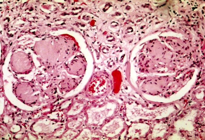
Any renal path experts here want to try to explain this a little better?
and for Jamiu: Yeah supposedly it would be more cellular with membranoproliferative if thats what you were referring to, so yeah, not enough black dots on that last one.
also, I know this was posted earlier in the thread but I think this might help people out:
proteus swarms on agar plates, two different ways it can be represented:


proteus swarms on agar plates, two different ways it can be represented:
newborn. ddx?
Blueberry muffin rash? Congenital CMV or rubella?
Similar threads


- Replies
- 0
- Views
- 727
