Extrapedicular Vertebral Augmentation

This forum made possible through the generous support of SDN members, donors, and sponsors. Thank you.

manowar rules

New Member
15+ Year Member
Joined
May 30, 2006
Messages
203
Reaction score
124
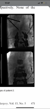
First time I have reason to do a true extrapedicular kypho. 60 y/o M on chronic glucocorticoids presents with severe low back pain. Edema noted at L3 and L4, patient with prior instrumented fusion at L4-5.

IMG_7078.jpg
IMG_7093.jpg
IMG_7094.jpg


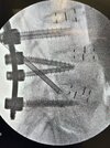
Screws are fairly superior at L4, so I was planning on using Beall's extrapedicular inferior end plate access, aka low and outside (3rd picture).
Since I hadn't augmented a fused level before, Stryker connected me to one of their KOLs. He was obviously an IR guy who did many of these under CT. He actually recommended that after augmenting L3, using the curved needle to go through the L3-4 disc and injecting cement into L4 that way. He also mentioned that if he gets into bleeding going extrapedicular, he will place in some Gelfoam or Surgicel. All this made me kind of nervous. I am but a simple country pain doc who does in-office kyphos under local and oral meds. Beall makes extrapedicular access sound easy, but I'm not trying to be a cowboy if this is some big ordeal. Any experience docs with thoughts or suggestions?

Members don't see this ad.
 
  • Like
Reactions: 1 user
He is crazy with that go through the disc talk. High and outside left side L3. High and outside right side L4. Please ask the Stryker rep to send me my customary $500.
 
  • Like
Reactions: 1 users
Members don't see this ad :)
He is crazy with that go through the disc talk. High and outside left side L3. High and outside right side L4. Please ask the Stryker rep to send me my customary $500.
Lol, glad to hear it. You don’t think I’d run into the pedicle screw going in high at L4? Do you agree with Doug about taking a 45 degree approach to enter lumbar extrapedicular?

Also, the boss rep recommended not inflating a balloon at the fused level but just doing vertebroplasty. Do you agree?

Your help and experience are very appreciated, thank you
 
Maybe about 38 degrees. 45 seems like too much. The trochar will just skip over the pedicle screw. You have to inflate the balloon. If the screw ruptures it, no big deal. The balloon is critical for making a nice pocket and keeping the cement contained, more ideally spread.
 
  • Like
Reactions: 1 user
I agree with Steve. Interested to what a radiologist calls it.

Most of my regular kypho I start extrapedicular, leads to a better midline balloon for me
 
  • Like
Reactions: 1 user
I agree with Steve. Interested to what a radiologist calls it.

Most of my regular kypho I start extrapedicular, leads to a better midline balloon for me
When you do your transpedicular approach, do you start off oblique and go coaxial to the pedicle or do you try to keep it more AP and come from the pedicle to the medial? Most IR guys I have seen drive down to pedicle in AP but I've seen both ways be done
 
Yes, agreed. Looks more like modic changes from the l3/4 adjacent segment disease.
I agree with this. Can you post more pictures, including T2 and stir?

I haven’t done vertebral augmentation since fellowship a decade ago… But I have done a dozen or so levels with extrapedicular access for intracept for adjacent fusion levels, as well as mid/upper lumbar levels where pedicle anatomy would not allow for access or posteromedial vert body targeting. In general, I actually find it easier and faster than transpedic, particularly at a level without pedicle screws. Navigating around the screw to then get to ideal targeting for intracept requires a few additional tricks, but without a screw at the level, it really is not bad at all. If you can do vert augmentation with traditional transpedicular access you can definitely do this.

My biggest points of caution would be to confirm on AP and lateral that you are definitely superolateral on pedicle, at junction with vertebral body, before you start malletting….. i.e. don’t go medial into the neural foramen before you are firmly in bone. Map out your path on MRI ahead of time. Don’t take a kidney biopsy, spear a nerve root or get more lateral on the vertebral body, where there are some big bad blood vessels.

I am submitting a technical report shortly on how to do this for intracept at adjacent levels to fusion. Technique has become pretty reliable after some initial troubleshooting.

Some pictures below accessing EP without fusion:
 

Attachments

  • 6689712B-7DAA-43C0-9D34-709C6E857AD4.png
    6689712B-7DAA-43C0-9D34-709C6E857AD4.png
    528.2 KB · Views: 76
  • 71157022-6E6C-41A7-8631-969B83B084C0.png
    71157022-6E6C-41A7-8631-969B83B084C0.png
    542.7 KB · Views: 77
  • 594A4168-CCEF-445C-B472-5E0403A8DECA.png
    594A4168-CCEF-445C-B472-5E0403A8DECA.png
    569.8 KB · Views: 78
  • FC0A60DB-1435-4217-B4F5-61B656DD432D.png
    FC0A60DB-1435-4217-B4F5-61B656DD432D.png
    527.9 KB · Views: 78
  • Like
Reactions: 3 users
Modic 1 superior adjacent segment disease. This is why we end up marching up levels after fusion. May become a spondy.

Also, WTF with the transdiscal approach? Those are words uttered exclusively by an individual who will never see that patient again, and will pawn him or her off onto someone else to manage his BS.
I came across his transdiscal approach a few months ago when I was sorting out how I was going to approach adjacent levels for intracept. Found them. Crazy? Balls? Genius? Uncertain…
 

Attachments

  • 35D42537-0E65-4791-AFE0-35C7C15AC77D.png
    35D42537-0E65-4791-AFE0-35C7C15AC77D.png
    188.4 KB · Views: 74
  • 58F19E89-C8E2-4383-B1B1-6E1A9092B644.png
    58F19E89-C8E2-4383-B1B1-6E1A9092B644.png
    202.7 KB · Views: 80
  • 0A17091F-9541-4057-924A-1FA8A81D7B4B.png
    0A17091F-9541-4057-924A-1FA8A81D7B4B.png
    187.7 KB · Views: 71
  • B167DA67-6B31-4090-9DC8-8BA491771D67.png
    B167DA67-6B31-4090-9DC8-8BA491771D67.png
    279.3 KB · Views: 76
  • CEE545AC-1076-4758-9965-A38E3820BD45.png
    CEE545AC-1076-4758-9965-A38E3820BD45.png
    249.5 KB · Views: 72
  • Like
Reactions: 1 user
Yeah you could be right. I don’t have access to the images this weekend but I’ll upload the STIR in a couple days. The radiologist called it “suspected acute/subacute L3 vertebral body compression fracture involving inferior endplate, approximate 10% height loss. Possible L4 superior endplate vertebral body compression fracture.” Interestingly it also mentions psoas muscle edema suggestive of acute strain or denervation. Not sure how I differentiate. Maybe if his worst pain is resisted straight leg raise?
 
Members don't see this ad :)
Yeah you could be right. I don’t have access to the images this weekend but I’ll upload the STIR in a couple days. The radiologist called it “suspected acute/subacute L3 vertebral body compression fracture involving inferior endplate, approximate 10% height loss. Possible L4 superior endplate vertebral body compression fracture.” Interestingly it also mentions psoas muscle edema suggestive of acute strain or denervation. Not sure how I differentiate. Maybe if his worst pain is resisted straight leg raise?
CT?
 
Yeah you could be right. I don’t have access to the images this weekend but I’ll upload the STIR in a couple days. The radiologist called it “suspected acute/subacute L3 vertebral body compression fracture involving inferior endplate, approximate 10% height loss. Possible L4 superior endplate vertebral body compression fracture.” Interestingly it also mentions psoas muscle edema suggestive of acute strain or denervation. Not sure how I differentiate. Maybe if his worst pain is resisted straight leg raise?
I wouldn't recommend you kypho that pt just yet. I'd shoot a bilateral TF and see how he does. CT may help. It looks like spondylotic effusion IMO. Our radiologists use that term a lot.

You MRI him in 12 months and there's prob fatty infiltrate and it's Modic 2.

Why the chronic steroid use?
 
I agree with this. Can you post more pictures, including T2 and stir?

I haven’t done vertebral augmentation since fellowship a decade ago… But I have done a dozen or so levels with extrapedicular access for intracept for adjacent fusion levels, as well as mid/upper lumbar levels where pedicle anatomy would not allow for access or posteromedial vert body targeting. In general, I actually find it easier and faster than transpedic, particularly at a level without pedicle screws. Navigating around the screw to then get to ideal targeting for intracept requires a few additional tricks, but without a screw at the level, it really is not bad at all. If you can do vert augmentation with traditional transpedicular access you can definitely do this.

My biggest points of caution would be to confirm on AP and lateral that you are definitely superolateral on pedicle, at junction with vertebral body, before you start malletting….. i.e. don’t go medial into the neural foramen before you are firmly in bone. Map out your path on MRI ahead of time. Don’t take a kidney biopsy, spear a nerve root or get more lateral on the vertebral body, where there are some big bad blood vessels.

I am submitting a technical report shortly on how to do this for intracept at adjacent levels to fusion. Technique has become pretty reliable after some initial troubleshooting.

Some pictures below accessing EP without fusion
 
@Taus I find your images more exciting than Tramsformers 2 and that is saying a lot! Well done!


You absolutely must start doing kyphoplasty. Intracept is much more difficult and you seem to have mastered it.
 
@Taus I find your images more exciting than Tramsformers 2 and that is saying a lot! Well done!


You absolutely must start doing kyphoplasty. Intracept is much more difficult and you seem to have mastered it.
Lol thanks!

How does it pay on pro fee in ASC? Wont be feasible in my office fluoro set up
 
The same as an Intracept.

But you can do it in any office you can do a ESI in. I have done them in a pretty pitiful space without issue.
 
A8CA53FA-3DE3-4FFA-A4A8-11AFD3DAB6D7.jpeg

Ok here are STIR and T2. Brightest thing is the disc, radiologist’s comment is simply “focal T2 hyperintense intervertebral disc signal.”
Yes my monitor is dusty.
 
I agree with this. Can you post more pictures, including T2 and stir?

I haven’t done vertebral augmentation since fellowship a decade ago… But I have done a dozen or so levels with extrapedicular access for intracept for adjacent fusion levels, as well as mid/upper lumbar levels where pedicle anatomy would not allow for access or posteromedial vert body targeting. In general, I actually find it easier and faster than transpedic, particularly at a level without pedicle screws. Navigating around the screw to then get to ideal targeting for intracept requires a few additional tricks, but without a screw at the level, it really is not bad at all. If you can do vert augmentation with traditional transpedicular access you can definitely do this.

My biggest points of caution would be to confirm on AP and lateral that you are definitely superolateral on pedicle, at junction with vertebral body, before you start malletting….. i.e. don’t go medial into the neural foramen before you are firmly in bone. Map out your path on MRI ahead of time. Don’t take a kidney biopsy, spear a nerve root or get more lateral on the vertebral body, where there are some big bad blood vessels.

I am submitting a technical report shortly on how to do this for intracept at adjacent levels to fusion. Technique has become pretty reliable after some initial troubleshooting.

Some pictures below accessing EP without fusion:
Nothing to fix there. Time for Nevro.
When you do your transpedicular approach, do you start off oblique and go coaxial to the pedicle or do you try to keep it more AP and come from the pedicle to the medial? Most IR guys I have seen drive down to pedicle in AP but I've seen both ways be done
start oblique but don’t go straight down the barrel, mark in a lateral to medial fashion just off the pedicle. Check spinal needle first in lateral and give local. After you engage you can just do AP/lat
 
  • Like
Reactions: 1 user
But… the cement will kill the basivertebral nerve just fine if we are being honest with ourselves.
 
  • Like
Reactions: 2 users
But… the cement will kill the basivertebral nerve just fine if we are being honest with ourselves.
I've thought about this. Truthfully, is there a difference between kypho and BVN ablation?
 
No, patients get pain relief after a bad structural fill due to killing the BVN. It would not take much cement to embolize the BVA.
 
  • Like
Reactions: 1 user
No, patients get pain relief after a bad structural fill due to killing the BVN. It would not take much cement to embolize the BVA.
So more people will call that schmorls node, or image above, a fracture and get paid more to cement it than burn it? Brilliant?!
 
  • Like
Reactions: 1 user
A transdiscal approach?? What??

I use a unilateral parapedicular approach above T10 usually. Never had to kypho a fused level. I have read about the inferior end plate approach and would be interested if anyone here has used it.

Map any approach out first on the MRI to avoid organs and nerves.

And lastly that looks like Modic change without a convincing fracture.
 
  • Like
Reactions: 1 user
Intracept got denied so call it a fracture
 
  • Haha
Reactions: 1 user
For one, this is why you should read your own MRI and not rely on radiologists reports
 
I would just pull back the trochar to the pedicle, or even into the pedicle if you are lateral, insert the beveled tip, yoke the handle lateral and drive the trochar medial to a new path and vertebroplasty if the cement was pooling too far anterior.
 
  • Like
Reactions: 1 user
What is your belief on this Steve? Fill as much as possible or fill every quadrant evenly and call it a day?
I believe old data where 2 to 4.5cc was enough to fix the fracture. Doug is gathering new data and I hope he publishes more than a textbook on the topic. He has experience beyond the rest of us combined and believes as complete a fill as possible to stabilize to fx is necessary. I cannot fill the bone as much as I see him do without getting extravasation somewhere.
 
  • Like
Reactions: 1 user

5-10cc depending on severity.


2-3.5cc

Lots of studies without agreement. Probably a % volume of fill would bo optimal to fill the fx and not increase adjacent level fx risk.
 
I believe old data where 2 to 4.5cc was enough to fix the fracture. Doug is gathering new data and I hope he publishes more than a textbook on the topic. He has experience beyond the rest of us combined and believes as complete a fill as possible to stabilize to fx is necessary. I cannot fill the bone as much as I see him do without getting extravasation somewhere.
Definitely have been a few times where more fill winds up with a bit of extravasating cement and I wish I would have stopped a second earlier. I also don’t see any difference in outcome so I’m inclined to the enemy of good theory.
 
  • Like
Reactions: 1 user
Do you guys have a hard cutoff on how high a kypho level you will do? Have a guy with 5 acute fractures... Two of them at T5 and 6. Haven't done any that high before. Any pointers?

Thanks
 
Do you guys have a hard cutoff on how high a kypho level you will do? Have a guy with 5 acute fractures... Two of them at T5 and 6. Haven't done any that high before. Any pointers?

Thanks
The longer you’re in the game to hire you will go. I’ve done a bunch of tea fives at this point in my career. When I was younger, I don’t think I would’ve ever gone above T8.
 
  • Like
Reactions: 1 user
This case kinda shocks me. What symptoms are you treating exactly? I’m with Steve Nevro for axial LBP or Epidural LOA for appendicular pain…
 
This case kinda shocks me. What symptoms are you treating exactly? I’m with Steve Nevro for axial LBP or Epidural LOA for appendicular pain…
He had sudden severe acute low back pain with twisting a couple weeks ago. I guess I should have emphasized the suddenness of onset. Minimal baseline pain, not on opiates. I called him yesterday and he actually was admitted to the hospital 2 days ago due to severity of pain.
 
Last edited:
I would just pull back the trochar to the pedicle, or even into the pedicle if you are lateral, insert the beveled tip, yoke the handle lateral and drive the trochar medial to a new path and vertebroplasty if the cement was pooling too far anterior.
Exactly what works well to stay posterior on EP intracept around fusion. Cranking it laterally with a bevel-tip stylet.
 

Attachments

  • 7DAA89B8-1A85-486B-B721-86E5787A9C2D.jpeg
    7DAA89B8-1A85-486B-B721-86E5787A9C2D.jpeg
    153.5 KB · Views: 66
  • F70E1419-8816-4B32-95AD-A3B132A5A5D0.jpeg
    F70E1419-8816-4B32-95AD-A3B132A5A5D0.jpeg
    198.5 KB · Views: 62
Top