Histopath images thread

This forum made possible through the generous support of SDN members, donors, and sponsors. Thank you.
Nevermind yaah, although myxoid type is one type of liposarc, the bluish hue is mucoid substance. The chickenwire vasculature gives it away and overall, the lesion looks low grade.

Members don't see this ad.
 
OK so this is an old guy who back in the day would get this really kickass "weed" from his dope dealer.
Fok-Yu: Yo yo yo. You got some good ****?
Hung-Man: I got some grade A weed here with THC crystals sprinkled on. It's called, "Starry sky", you gotta try some of this **** out!
Fok-Yu: How much for an eighth?
Hung-Man: Three-fiddy.
Fok-Yu: What a bargain! I don't even need coupon. OK, gimme five of them.

What Fok-Yu didn't realize was that Hung-Man was an evil evil man and those weren't THC crystals sprinkled on that weed!

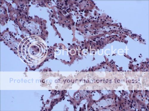
Many years later, Fok-Yu has to undergo an extrapleural pneumonectomy cuz he got some bad **** all over his lung!

wonton.jpg


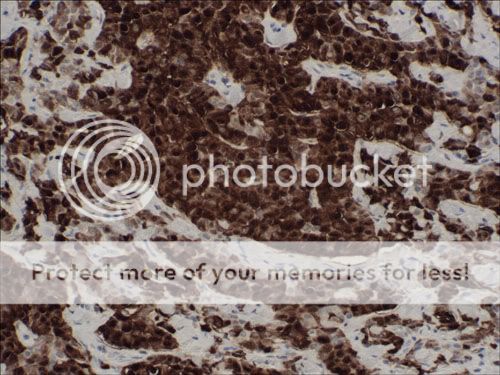
The tumor cells stained positive for calretinin...

wonton_Calretinin.jpg


...and they stained positive for WT-1...

wonton_WT-1.jpg


So what is Fok-Yu's diagnosis?
 
...but see, Hung-Man had a reason to be evil. See, back in the day, Hung-Man was angry at the world. Hung-Man really wasn't a man...he was a woman but in her late 30's, Hung-Man started to look like a man cuz she initially grew a mustache but then it became a fullblown beard. Hung-Man was used to shaving her legs to look sexy for the men but Hung-Man would now have to start shaving her face. Hung-Man very very angry. Hung-Man felt that God had cheated her out of a good life. Well one day, Hung-Man was diagnosed with a pretty sizable adrenal mass...and this is what it showed:

At lower power (for the higher minds in the audience):
stirfry_low.jpg


At higher power (for those who want to refine their diagnosis), you had two areas of histology that were of interest:

stirfry_high_1.jpg


and

stirfry_high_2.jpg


Why does Hung-Man suddenly look like Santa Claus with a *****?
 
Members don't see this ad :)
AndyMilonakis said:
spell out MPM and I might give you credit :)

muh patchy mungs?
:)

malignant pleural mesothelioma? :eek:
 
quant said:
muh patchy mungs?
:)

malignant pleural mesothelioma? :eek:
You got it. The force is strong in you. You have a high metaclorian index (Star Wars started to suck when they introduced this concept, IMHO).

Can you guess what Hung-Man has?

Hint: Hung-Man never complained of palpitations or headaches.

BTW, this mass did NOT stain positive for S100, chromogranin, or synaptophysin. OK, now your differential is now reduced to 3 things instead of 4 :)
 
AndyMilonakis said:
Hint: Hung-Man never complained of palpitations or headaches.

BTW, this mass did NOT stain positive for S100, chromogranin, or synaptophysin. OK, now your differential is now reduced to 3 things instead of 4 :)
1) Adrenocortical carcinoma of some sort
2) Ovarian sex cord stromal tumour
3) ...dunno... lack of midiclorians?

Andy go home!
 
AndyMilonakis said:
You got it. The force is strong in you. You have a high metaclorian index (Star Wars started to suck when they introduced this concept, IMHO).

Can you guess what Hung-Man has?

Hint: Hung-Man never complained of palpitations or headaches.

BTW, this mass did NOT stain positive for S100, chromogranin, or synaptophysin. OK, now your differential is now reduced to 3 things instead of 4 :)


Some kind of CAH?

But the age of onset excludes it. No palpitations or headaches, pheochromocytoma is out.

No stain of chromogranin?....neuroendocrinal cells are out, unless this is some wicked variant.

Dunno, id still say some kind of CAH... :eek: :eek:
 
mcfaddens get the prize.

Although adrenal cortical carcinomas are quite rare, this tumor had several features consistent with it:

1. Capsular invasion (not shown in above pics)
2. Atypical mitoses
3. Over 5 mitoses per 50 hpf. In this tumor, there were over 20 mitoses per 50 hpf (40X). In fact, you can spot 3 bizarre mitotic figures in one 20X field in that picture.
4. Lymphovascular invasion (which was present but not shown in the pics above).
5. High Fuhrman nuclear grade (in this tumor, we decided that the Fuhrman grade was III-IV out of IV...I was leaning towards IV but that's moot. Fuhrman grade of III helps you put this tumor in the malignant category).
6. Necrosis (which was present in the last pic).
7. Sheetlike arrangement of cells in which 75% or more of the cells have prominent eosinophilic cytoplasm.

The history (hirsutism) isn't specific for the diagnosis as this could still suggest a virulizing adrenocortical adenoma. But when most people think of nasty looking adrenal tumors, people tend to think of pheochromocytoma first which is typically categorized by headaches, palpitations, paroxysmal hypertensive episodes, etc.

quant and deschutes, your clinical reasoning was quite nice.
 
quant said:
MORE MORE!!!!
Trust me. I will be putting up more cool cases as I see them. This week I was on biopsies...so lots of bread and butter stuff but nothing mind-blowing. I always take pictures of any cool cases when I encounter them so as I have time, I'll post some of them here. There is one really freaky case which I don't feel comfortable posting because there may be a slight possibility that it will get written up.

To the other residents ... if you can, please post some cool cases. We can all learn a lot from each other. :thumbup:
 
OK apparently deschutes is throwing a hissy fit cuz she had the correct answer in her differential list. I had misread her post. But nonetheless, she didn't commit to a single answer. So she gets no cookie.

Yeah and then deschutes is like, but ACC could mean adenoid cystic carcinoma. But this is no adenoid cystic carcinoma. And I think when mcfaddens said ACC, he or she (no clue if mcfaddens is a proud owner of a penis or not) didn't mean this.
 
Methinks thou doth protesteth too much :p

AndyMilonakis said:
OK, now your differential is now reduced to 3 things instead of 4 :)
This is the reason I had 3 items on my list, with "adrenocortical carcinoma of some sort" at the top.

What are the other 2 differentials (or three, if you will)?
 
deschutes said:
Methinks thou doth protesteth too much :p
Methinks you need to quit yo bitchin'! ;)
This is the reason I had 3 items on my list, with "adrenocortical carcinoma of some sort" at the top.

What are the other 2 differentials (or three, if you will)?
When I think of adrenal neoplasms, I think of 4 main things:
1. Adrenal adenoma (I guess hyperplasia too...but that's not exciting)
2. Adrenal cortical carcinoma
3. Pheochromocytoma
4. Metastasis
 
Members don't see this ad :)
AndyMilonakis said:
Methinks you need to quit yo bitchin'! ;)
But you take it so well... :D

AndyMilonakis said:
When I think of adrenal neoplasms, I think of 4 main things:
1. Adrenal adenoma (I guess hyperplasia too...but that's not exciting)
2. Adrenal cortical carcinoma
3. Pheochromocytoma
4. Metastasis
So what does...

this mass did NOT stain positive for S100, chromogranin, or synaptophysin.
...rule out?

synaptophysin stains neuroblastoma, that's all I know.
 
AndyMilonakis said:
rules out pheo.

whoa slow down guys....
at this rate i won't have anything left to learn in the residency!!!! :laugh: :laugh:
 
quant said:
whoa slow down guys....
at this rate i won't have anything left to learn in the residency!!!! :laugh: :laugh:
ok you've had the weekend off :) more to come...just gotta take the pics.

anyways, one can never see too much stuff before residency. i wish i could've seen more histopath before starting...but that's partly my fault because i really didn't take the initiative to do so.

the number of times i've seen a slide while previewing and thought, WTF...countless.
 
quant said:
at this rate i won't have anything left to learn in the residency!!!! :laugh: :laugh:
No such thing!!

On the other hand, I still can't convince myself that I lost anything by not doing a PSF. I mean, 20 years out no one is going to care if you did a PSF or not.
 
This happens to me every day:

Me: What's that?
Attending: What's what?
Me: The area I dotted
Attending:I have no idea, but it isn't significant.
 
yaah said:
This happens to me every day:

Me: What's that?
Attending: What's what?
Me: The area I dotted
Attending:I have no idea, but it isn't significant.
Yeah. I've practically limited my painstaking circling to elusive blasts and Auer-rodded blasts.

~
My bone marrow biopsy patient this morning kept farting. I think I smelt beans and onions... :mad: :laugh: :laugh:
 
yaah said:
This happens to me every day:

Me: What's that?
Attending: What's what?
Me: The area I dotted
Attending:I have no idea, but it isn't significant.
It feels good when you get a consult slide and in the report, the previous pathologists missed a finding...like a positive lymph node! Holla!

Of course, at signout, I don't say, "Here's a lymph node they missed." Instead, I'm like, "I dotted a suspicious area in a lymph node...what do you think about this?"
 
I haven't seen a case of adenoid cystic carcinoma (or whatever that ACC entity was called) but when I do, and I get pimped on it, I can confidently recognize it. Then when they ask me how I knew that, I'll be like, "Cuz I'm the f*cking bomb! Recudnize!"

Today, I saw some other cases that I haven't posted yet so hopefully I'll have some time this weekend to take some more photos and post them here.
 
Guy goes into see his doctor..."Something's wrong with me. I **** 100 times a day. It's wrong."

Doctor orders a CT scan...at the radiology suite, the guy is told to stay still. The guy has to fight the urge to **** his pants. Finally, the CT scan is over and he runs to the bathroom to land a steaming dump.

Later, he returns to his doctor. Doctor says, "I've got some bad news and some good news."

"What's the good news?" asks the patient.
"There's nothing wrong with your ass."
"What's the bad news?" retorts the patient.
"There's a mass in your kidney...we're gonna have to jam a needle in your side and take a biopsy. Yup. It'll take a little while, you're gonna have to fight the urge to crap your pants yet again."

This is the biopsy specimen.

BloodPisser.jpg
 
A lady comes into the ER. When asked about her chief complaint, she squeamishly responds, "Um...there's something wrong down there."

One thing leads to another and they do an X-ray. They find this:
daaaaaamn.jpg


OK well that's all fine and dandy as she explains, "Uh yeah I accidentally fell on it." ER physician is like, "Yeah whatever...oh btw, I noticed you were 51 years old, have you had a mammogram recently?"

"No Doctor, I've never had one of those."

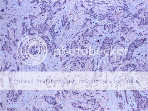
Well, later the mammogram is completed, they see a mass, they cut off her boob, and wah-lah!

In one area, you see this:
Titty1.jpg


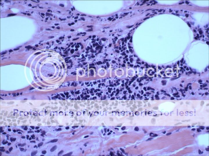
In an adjacent area, you see this:
Titty2.jpg
 
AndyMilonakis said:
man, i just saw a carcinoid case and was gonna put it up. well, lo and behold, what do i see, the last post in this thread deals with carcinoid.

ok, well, there's that case going out the sh*tter.

here's another cancer case. also involving the appendix like your carcinoid case. btw, this tumor was part of a bigger colon resection too. i don't recall exactly where the primary is (came from an outside hospital and we don't have history) but it's likely to be somewhere from the GI tract as it was Ck20 and CDX2 positive.

enjoy!
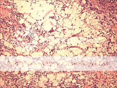
AssCancer.jpg


And here are the Ck20 and CDX2 stains, in respective order.
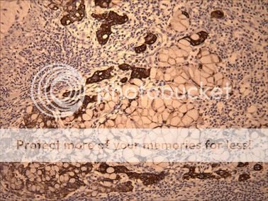
CK20.jpg

CDX2.jpg

Addendum: deschutes is throwing yet another hissy fit! "what's the answer? what's the answer? wah wah wah...i want my mommy!"

OK the answer is signet cell adenocarcinoma.
 
yaah said:
I dunno - papillary RCC?

I have to take some pictures of the vag specimens I saw this week - some crazy shi in there.
Word up dude, it is papillary RCC.

Interestingly, when we got the frozen specimen, it looked like an oncocytoma since the tumor cells were very eosinophilic without perinuclear halos. Of course, we didn't have a good sense of the architecture of the specimen until we received more of the tumor specimen for permanents.

Well done. you get a cookie.

Now, go sacrifice some lambs so that the football gods smile on the wolverines tomorrow when they face the f*cking gophers.
 
OK. There's this guy. Whenever he's on bottom, he gots this weird chest pain. When he be doin' her doggy-style, no chest pain.

Long story short...dude ended up getting a pericardial biopsy and this is what you saw.

Low power:
endgame1.jpg


Two High power shots:
endgame2.jpg


endgame3.jpg
 
Chief complaint: Difficulty swallowing.
HPI: 39 yo female comes in complaining that she has progressive dysphagia. It is ruining her sex life. She is accompanied by her husband of 12 years who states that it would be nice if she could swallow after she does the you know what.
Past Surg Hx: Thyroidectomy
Physical exam: Multiple nodular masses noted on the right neck.
A/P: Right radical neck dissection

Here are some pics of one of the lymph nodes. 40 of 60 nodes had this.
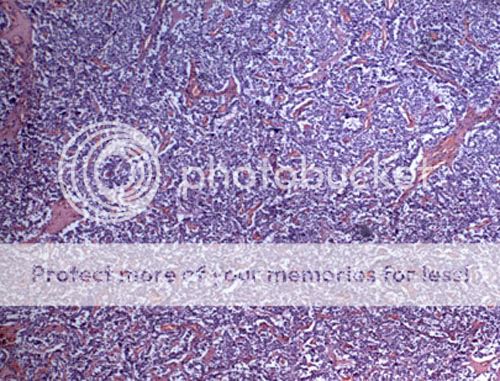
Low power (sorry for the sh*tty focus):
spitorswallow1.jpg


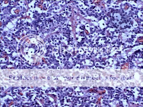
High power:
spitorswallow2.jpg
 
Pericarditis for the first. I thought of adding on myocarditis, but I don't understand why the myocardium has got fat in it.

The second, I'm going to go for broke and say it's metastatic i.e. non-lymphoid/hematopoietic. I see two cell populations (large clear cells with small round dark nuclei, and smaller basophilic cells with more open chromatin).

Otherwise clueless ;)
 
You're right deschutes. It was metastatic. It happened to be small cell carcinoma. And some of the morphological features are consistent with the diagnosis: single cell necrosis, molding, nuclear pleiomorphism, and mitoses. In fact, a lot of small cell carcinomas can cause a smudging kind of artifact on frozen sections and permanents. Not a pathognomonic feature since lymphoid cells can smudge too.

It could be some kind of lymphoma and that is certainly on the differential. In retrospect, I should have put down that the tumor cells were positive for Ck7 and TTF-1 which is consistent with a tumor of pulmonary origin.

The clinical history was a distractor...on purpose. In fact, the sex part was made up but the pericarditis-like presentation was true. But we diagnose based on morphology, not clinical presentation!
 
AndyMilonakis said:
It happened to be small cell carcinoma. And some of the morphological features are consistent with the diagnosis: single cell necrosis, molding, nuclear pleiomorphism, and mitoses.
I'm staring at the high-power. Are ANY of those features (apart from perhaps nuclear pleomorphism) in that field :confused:

AndyMilonakis said:
The clinical history was a distractor...on purpose.
"Distractor"? I thought it only helped! Positional chest pain ;)
 
deschutes said:
I'm staring at the high-power. Are ANY of those features (apart from perhaps nuclear pleomorphism) in that field :confused:
It's there--some stuff more apparent than others. In fact, I was able to spot it some of it at low power ;)

"Distractor"? I thought it only helped! Positional chest pain ;)
but it's not pericarditis. those aren't inflammatory cells. fine fine...you caught me...it was a trick question.
 
AndyMilonakis said:
It's there--some stuff more apparent than others. In fact, I was able to spot it some of it at low power ;)

but it's not pericarditis. those aren't inflammatory cells. fine fine...you caught me...it was a trick question.

So, pericardial lymphoma then?
 
AndyMilonakis said:
The case was signed out as metastatic small cell carcinoma.

What about the first one? They were both SCC? Please forgive my density.
 
Aubrey said:
What about the first one? They were both SCC? Please forgive my density.
You don't need to ask me for forgiveness...that's between you and Lord Virchow.

OK so I put up 2 cases. The first dude with the chest pain when in a certain sex position...that was metastatic SCC.

The second case...the spit can't swallow no more lady...the answer to that was medullary thyroid carcinoma in a lymph node (which became a problem for her a few years after her initial thyroidectomy).
 
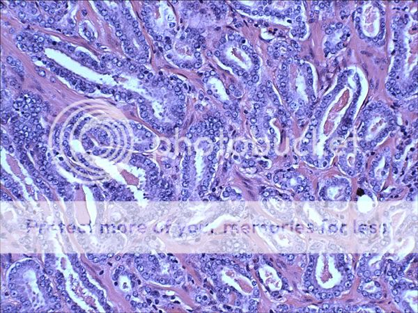
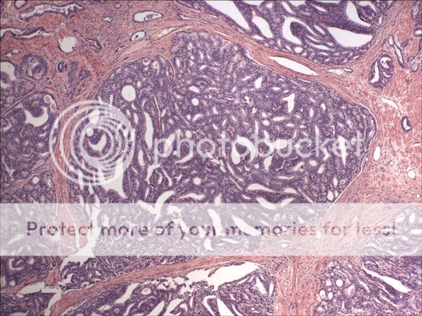
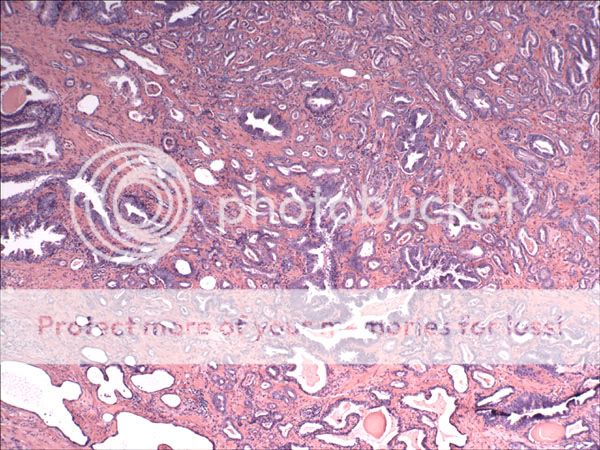
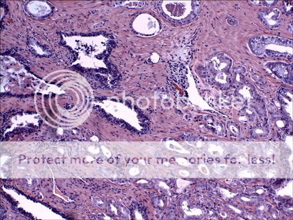
Here's a patient who had a really nasty prostatic adenocarcinoma...various shots from various sections:
nutgobbler1.jpg

nutgobbler2.jpg

nutgobbler3.jpg


with a focal component of ductal adenocarcinoma:
nutgobbler4.jpg
 
deschutes said:
24 y/o girl gets a hernia repair. Few weeks later, voila! Another lump in the same spot. FNA shows:

U05-6884_10x_1.jpg

U05-6884_10x_2.jpg

U05-6884_50x_1.jpg

U05-6884_50x_2.jpg

Is that a core bx. ive never seen a FNA look like that.

It looks like something poorly diffrentiated, maybe some epithelial neoplasm,
 
mcfaddens said:
Is that a core bx. ive never seen a FNA look like that.

It looks like something poorly diffrentiated, maybe some epithelial neoplasm,
That was my thought too...but you would need history to see if there has been any documented primary (assuming this is a metastasis in soft tissue). If that was not available, impox studies would be fruitful...hopefully.
 
A panel of immunoperoxidase stains shows the cells to be positive for ALK, CD30, EMA, CD45, CD5, and possibly weakly for CD43. They are negative for CD3, CD20, cytokeratin AE1/AE3 and CA-125.
 
Necrotizing Lymphadenitis - Cat Scratch, tularemia, yersinia, or chlamydia and if not infectious then maybe Kawaski (sp?) however its got a lot of eos in there so I also have Hodgkin's in my differential.
 
deschutes said:
A panel of immunoperoxidase stains shows the cells to be positive for ALK, CD30, EMA, CD45, CD5, and possibly weakly for CD43. They are negative for CD3, CD20, cytokeratin AE1/AE3 and CA-125.

Generally ALK positivity means anaplastic lymphoma. Plus, aren't you on heme path? ;)

I'm gonna have to go with Hodgkin Lymphoma on #2.
 
yaah said:
Generally ALK positivity means anaplastic lymphoma. Plus, aren't you on heme path? ;)
I'm actually "off" hemepath :p But you're right, like I said to Andy, hemepath is the closest to AP that I've been so far.

Yup, the first case was signed out as Anaplastic Large Cell Lymphoma.

Neck node was Nodular Sclerosis Hodgkin Lymphoma. (2nd pix has my token representation of "fibrous bands", and you can see lacunar cells in the 3rd pix.)

IHC was positive for CD15, CD30, and negative for CD45. Which is classic.

Here is a nice CD30:
USR05-752_CD30_2.jpg
 
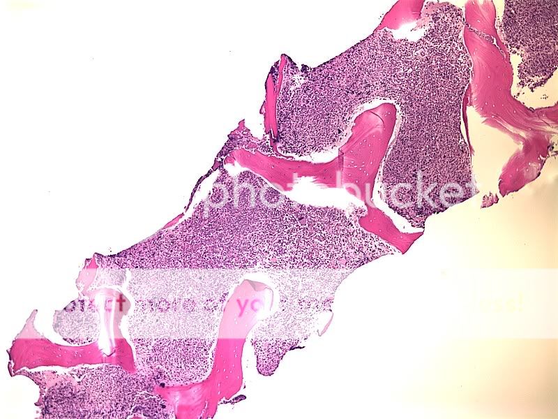
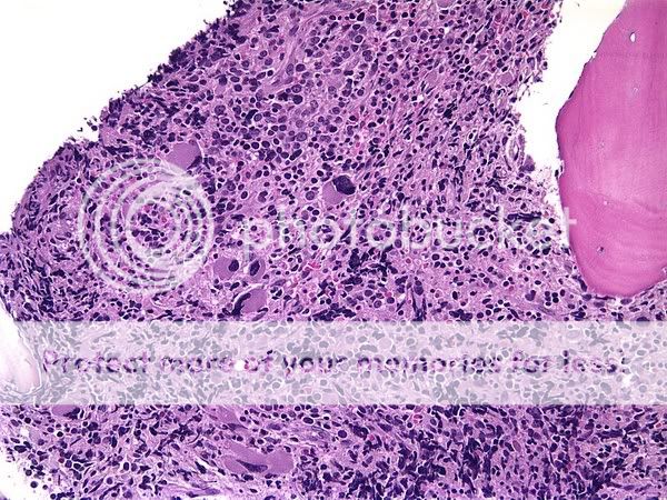
9 year old with a history of sarcoma (MPNST with rhabdoid cells - a.k.a. Triton Tumor) now with pancytopenia - bone marrow bx

28ab69fc.jpg

74b845fa.jpg

17de1653.jpg
 
So um, those are metastatic tumour cells?

I am continually surprised by how large the rest of the cells of the body are, in comparison with hematopoietic cells (megakaryocytess excluded). I mean, surg path scopes don't have 100x objectives!
 
That's the differential, and why it's an interesting case. Are those metastatic tumor cells in the marrow, or are they megakaryocytes (regular ones or dysplastic ones)? And either way, what is up with the rest of the marrow? Normal tumor replaced marrow becomes fibrotic, and this is mostly packed with cells and not fibrosis.

In short, this was a case for immunostains. We ordered so many immunostains this week that the attending was saying today, "Let's order Kappa Lambda, CD20, Cd3, CD10, and oh how about CD1a, and stop me before I order any more." So this one was stained for the markers that stained the original tumor cells (Myogenin, Desmin, etc) as well as markers for Megas and hematopoeitic elements to determine if all these cells were dysplastic red cells or what. Peripheral smear apart from pancytopenia was normal (aka no blasts) except for some teardrop red cells.
 
Top