
You are using an out of date browser. It may not display this or other websites correctly.
You should upgrade or use an alternative browser.
You should upgrade or use an alternative browser.
Pictures of the Week
Started by Epidural

Get help with your application
Use all the free resources available to you from SDN: articles, guides, expert advising, forums discussions, and school research.
Thanks for sharing this! I hadn't seen it.
This study did suggest that 5 nerves (adding IPBSN and recurrent fibular) might work better than 3, but the improvement seems so marginal that I don't know if it's worth it. Comparing effectiveness of 3-needle approach versus 5-needle approach of genicular nerve block on pain and quality of life in chronic osteoarthritis of knee: a double blinded randomised controlled trial - PMC
I personally really love this study of 15 cadavers by Peng, where he created a heat map of the genicular nerves. Anatomical Study of the Innervation of Anterior Knee Joint Capsule: Implication for Image-Guided Intervention - PubMed
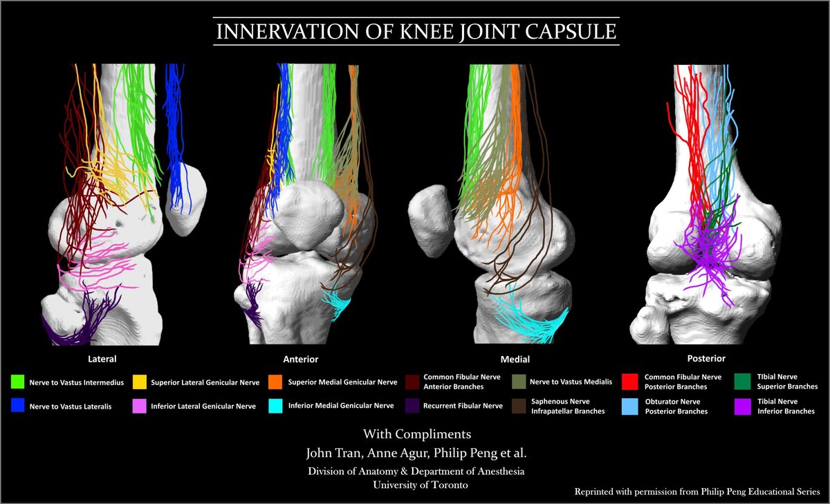
I think the infrapatellar branch has way too much variability to reliably RF, and it's so superficial I'm afraid of lesioning the skin. The superior lateral genicular nerve seems to have a lot of variability too, so I've been doing either bipolar or a second lesion for that spot.
I've personally found genicular RF to be among my highest satisfaction procedures.
I also wonder if ultrasound my be more accurate or provide better results. Did one case under ultrasound and placed the probe about 1 cm proximal to the corresponding genicular arteries with great results. Too much of a pain in the butt under ultrasound, however.
This study did suggest that 5 nerves (adding IPBSN and recurrent fibular) might work better than 3, but the improvement seems so marginal that I don't know if it's worth it. Comparing effectiveness of 3-needle approach versus 5-needle approach of genicular nerve block on pain and quality of life in chronic osteoarthritis of knee: a double blinded randomised controlled trial - PMC
I personally really love this study of 15 cadavers by Peng, where he created a heat map of the genicular nerves. Anatomical Study of the Innervation of Anterior Knee Joint Capsule: Implication for Image-Guided Intervention - PubMed
I think the infrapatellar branch has way too much variability to reliably RF, and it's so superficial I'm afraid of lesioning the skin. The superior lateral genicular nerve seems to have a lot of variability too, so I've been doing either bipolar or a second lesion for that spot.
I've personally found genicular RF to be among my highest satisfaction procedures.
I also wonder if ultrasound my be more accurate or provide better results. Did one case under ultrasound and placed the probe about 1 cm proximal to the corresponding genicular arteries with great results. Too much of a pain in the butt under ultrasound, however.

I wouldn't ever target recurrent fibular nerve given the obvious risks associated with fibular nerve being in proximity.Thanks for sharing this! I hadn't seen it.
This study did suggest that 5 nerves (adding IPBSN and recurrent fibular) might work better than 3, but the improvement seems so marginal that I don't know if it's worth it. Comparing effectiveness of 3-needle approach versus 5-needle approach of genicular nerve block on pain and quality of life in chronic osteoarthritis of knee: a double blinded randomised controlled trial - PMC
I personally really love this study of 15 cadavers by Peng, where he created a heat map of the genicular nerves. Anatomical Study of the Innervation of Anterior Knee Joint Capsule: Implication for Image-Guided Intervention - PubMed
View attachment 413645
I think the infrapatellar branch has way too much variability to reliably RF, and it's so superficial I'm afraid of lesioning the skin. The superior lateral genicular nerve seems to have a lot of variability too, so I've been doing either bipolar or a second lesion for that spot.
I've personally found genicular RF to be among my highest satisfaction procedures.
I also wonder if ultrasound my be more accurate or provide better results. Did one case under ultrasound and placed the probe about 1 cm proximal to the corresponding genicular arteries with great results. Too much of a pain in the butt under ultrasound, however.
I also second success/patient satisfaction with genicular RFA. It has to be effective since it may be the most painful procedure I perform, and patients keep coming back...
Advertisement - Members don't see this ad
Definitely never do anything you're not comfortable with!
I've never done personally targeted the recurrent fibular, but it seems reasonable for patients who greatest pain is anterior, or especially anterolateral and inferior.
If you're hitting the anterior tibia at the lateral aspect of its cortex, at the level of the fib head, you'd be well away from the peroneal nerve, which is still posterior and hasn't reach the fib neck yet (check it out on ultrasound!).
I've never done personally targeted the recurrent fibular, but it seems reasonable for patients who greatest pain is anterior, or especially anterolateral and inferior.
If you're hitting the anterior tibia at the lateral aspect of its cortex, at the level of the fib head, you'd be well away from the peroneal nerve, which is still posterior and hasn't reach the fib neck yet (check it out on ultrasound!).
Yeah I do standard for one burn, modified for second burn. So much anatomic variability that even doing that with 18 ga sidekick I'm skeptical that I'm really hitting themJokes aside, has anyone else read this paper?
It shows different targets than classically taught (to be clear, the pic above is terrible by classical or this paper's standards)
Curious what people think

Agree, I find people are generally very happy with results but it definitely seems to hurt like hell for most people. Did one today and the lady was miserable despite 3cc of 2% lido down each cannula for a full 2 min prior.I wouldn't ever target recurrent fibular nerve given the obvious risks associated with fibular nerve being in proximity.
I also second success/patient satisfaction with genicular RFA. It has to be effective since it may be the most painful procedure I perform, and patients keep coming back...
Anyone have any magic ways of making it hurt less?
I’ve tried lots of things. Lately I infiltrated 1% lido slowly on the way in with the RF needle, then get to target depth, pull back a cm and inject 2% lido as I push it back in. Seems to help but some still have trouble.Agree, I find people are generally very happy with results but it definitely seems to hurt like hell for most people. Did one today and the lady was miserable despite 3cc of 2% lido down each cannula for a full 2 min prior.
Anyone have any magic ways of making it hurt less?

I try not to address these branches unless absolutely necessary. I hit this less than 10% of the time and wouldn't recommend without ultrasound. You can usually find some small branches that are far medial and superior to the common peroneal nerve traversing inferior/lateral to the fibular head. In general, I'm far more concerned with how superficial these nerves are (and close to the joint capsule in TKA cases) than how close to the common peroneal they are.I wouldn't ever target recurrent fibular nerve given the obvious risks associated with fibular nerve being in proximity.
I also second success/patient satisfaction with genicular RFA. It has to be effective since it may be the most painful procedure I perform, and patients keep coming back...

Not seen by me. Rheumatology (partner) was consulted for neck/b/l arm pain from Neurosurgery. MRI with mild DDD and mild canal stenosis C5-6, C6-7.
These Xrays done 2 months earlier. Patient reports a fall hitting head. Neg MRI brain or T-spine. MRI's done to r/o MS as + Lhermitte's. Xray report read as negative. Guess why I am posting?
I'll give it a shot. Flex/ex XR shows a grade 1 anterolisthesis most prominent at C4 on C5. Sadly, could be a missed SCI around that level from acute cervical extension leading to a central cord syndrome.View attachment 413757
Not seen by me. Rheumatology (partner) was consulted for neck/b/l arm pain from Neurosurgery. MRI with mild DDD and mild canal stenosis C5-6, C6-7.
These Xrays done 2 months earlier. Patient reports a fall hitting head. Neg MRI brain or T-spine. MRI's done to r/o MS as + Lhermitte's. Xray report read as negative. Guess why I am posting?

I have the local anesthetic for genicular RFA figured out. The key is to put the c arm in lateral and take a local needle from lateral to medial to the area you target and drop in 1-2ml .5% ropi. It takes less than a minute to do all 3. Then you go to AP, 1% lido for the skin and as deep as the local needle will go. Place your rf needles. 2% lido through the rf needles. I then get the probes out and let the MA test motors. I know it isn’t needed but we are just killing time and they aren’t capable of knowing when or when not to test motors. Then lesion. They went from crying out to chilling after I added the initial ropi. It probably has 8 minutes before you start ablating to work.
I also do my blocks in lateral with this same approach.
there isnt significant soft tissue swelling and that anterolisthesis is minor i can see why rads called it normal.
the protocol i used for neck injury after fall with negative xrays and persistent pain is to get an MRI scan in ER or to send home patient home in a cervical collar and get MRI scan 4 weeks after.
the protocol i used for neck injury after fall with negative xrays and persistent pain is to get an MRI scan in ER or to send home patient home in a cervical collar and get MRI scan 4 weeks after.
She had MRI. C5-6 and C6-7 mild stenosis, no listhesis at C4-5.there isnt significant soft tissue swelling and that anterolisthesis is minor i can see why rads called it normal.
the protocol i used for neck injury after fall with negative xrays and persistent pain is to get an MRI scan in ER or to send home patient home in a cervical collar and get MRI scan 4 weeks after.
But there is 3mm slip in flexion and 0mm in extension.
Instability defined as: 2mm flex/ext or 3.5mm static.
so you think that explains radicular neck and arm pain? (you didnt comment about motor weakness or sensory loss...)
theres no listhesis and mild spinal stenosis on MRI scan and no T2 changes, so doesnt sound like central cord syndrome due to the injury.
what is your plan?
theres no listhesis and mild spinal stenosis on MRI scan and no T2 changes, so doesnt sound like central cord syndrome due to the injury.
what is your plan?
Advertisement - Members don't see this ad
there is a lot of ROM with this woman. you will probably see some amount of instability when there is that big of range on flex/ext. not surgical.She had MRI. C5-6 and C6-7 mild stenosis, no listhesis at C4-5.
But there is 3mm slip in flexion and 0mm in extension.
Instability defined as: 2mm flex/ext or 3.5mm static.
My impression is neck pain with bilateral arm pain means rule out central etiology (ie dynamic instability causing central canal compression). @lobelsteve mentioned +Lhermittes which prompted MRI to r/o spinal injury.so you think that explains radicular neck and arm pain? (you didnt comment about motor weakness or sensory loss...)
theres no listhesis and mild spinal stenosis on MRI scan and no T2 changes, so doesnt sound like central cord syndrome due to the injury.
what is your plan?
Definitely would expect T2 weighted changes. Usually, MRI only misses this in the immediate post injury setting before edema sets in (first few hours)Central cord should reveals something on the MRI right? Signal change?
@lobelsteve curious if she has UE weakness or subtle dexterity issues

I think you're wrong here Steve. Instability is defined as 3.5mm on flex/ext, not static flims. When did this change?She had MRI. C5-6 and C6-7 mild stenosis, no listhesis at C4-5.
But there is 3mm slip in flexion and 0mm in extension.
Instability defined as: 2mm flex/ext or 3.5mm static.
it hasn't 3.5mm on F/E, or 11 degree angular change-> instability
Medicare Administrative Contractors (MACs) use the below definition, hence using the below numbers:
3.5mm on F/E, or 11 degree angular change-> instability
I am not saying that lesser numbers could not be, I am just saying thats where they arbitrarily drew the line
3.5mm on F/E, or 11 degree angular change-> instability
I am not saying that lesser numbers could not be, I am just saying thats where they arbitrarily drew the line
so you are sending her for c4/5 ACDF?
Don’t think it is going to happen.
Don’t think it is going to happen.
Again: never saw patient. Looked at xray for colleague and saw listhesis but normal report. Then given history of neck trauma.
I’ve seen lots of clefts before in compression fractures, but don’t think I’ve seen this much air on the x-ray, including AP. 8cc densely filled in that void.
Attachments


A little long in the tooth?Patient in room 13 will do well.View attachment 414001
Nice glasses though.
CESI easy as they opened it wide between the lamina.

Very old. She doesn’t have X or Facebook.A little long in the tooth?
Nice glasses though.
CESI easy as they opened it wide between the lamina.
I like the glasses, too! May not have teeth, but still has style!
Advertisement - Members don't see this ad
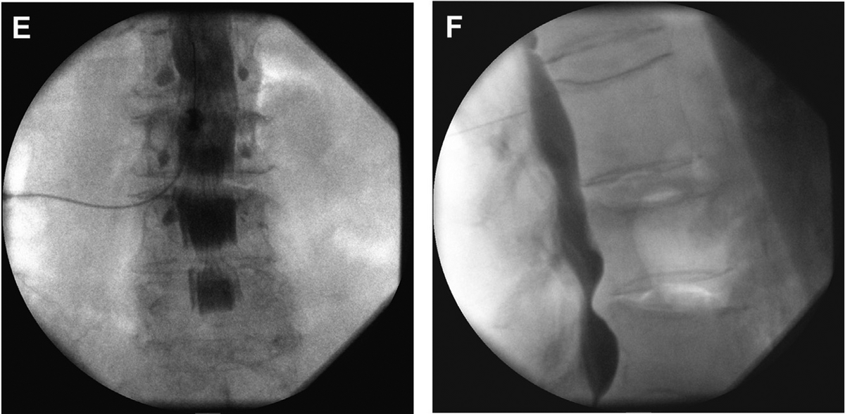
Not IT. Too far anterior and not medial enough with needle. And sheathogram.
Last edited:
Looks fine to me. I don't think that's too anterior at all. I shoot for anterior half of foramen.
Yeah, nice nerve root sheath with wrapping around from the ventral space is my guess. Get a STAT CT next time so I can know if I'm right?Intrathecal?
Contrast crossed over and epidural fat blobs are visible.
Thought that was odd, pulled back far enough I am lateral in the foramen and mostly getting nerve root pattern and it still crossed midline.
Got a lateral.
Aspiration negative for CSF.
Am I missing something? Contralateral spread is very normal for TFESI and, if anything, probably suggests good ventral spread into the epidural space. 6 mL of injectate has been shown to spread contralateral in most patients (Comparison of Epidural Spreading Patterns and Clinical Outcomes of Transforaminal Epidural Steroid Injection with High-Volume Injectate via the Subpedicular Versus the Retrodiscal Approach - PubMed) though I'm sure it eventually spreads contralateral with smaller volumes, assuming the injection is epidural and not just nerve root.
Am I missing something? Contralateral spread is very normal for TFESI and, if anything, probably suggests good ventral spread into the epidural space. 6 mL of injectate has been shown to spread contralateral in most patients (Comparison of Epidural Spreading Patterns and Clinical Outcomes of Transforaminal Epidural Steroid Injection with High-Volume Injectate via the Subpedicular Versus the Retrodiscal Approach - PubMed) though I'm sure it eventually spreads contralateral with smaller volumes, assuming the injection is epidural and not just nerve root.
I don’t think I can tell you the last time I saw contralateral spread off a TF. It was crossing over with 0.2cc of contrast. Like, first few drops crossed over.
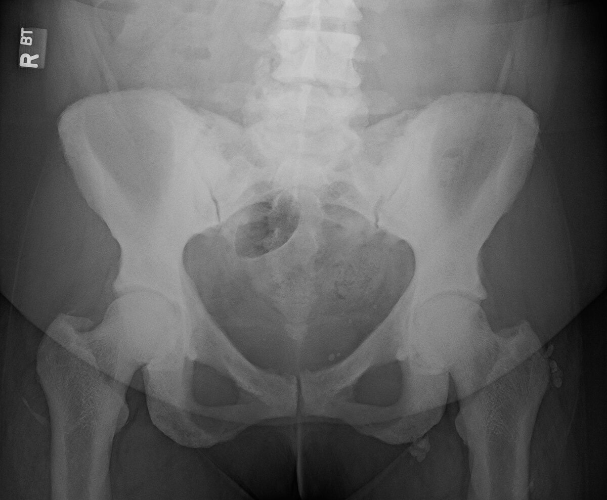
Osteopetrosis? MRI L spine with very dark bones on T1 and T2. Patient was sent for consult regarding Intracept, and she does have some Modic changes, but I’m very hesitant about the idea of trying to get through that bone. Sending her to endocrinology for a consult.
Osteopetrosis? MRI L spine with very dark bones on T1 and T2. Patient was sent for consult regarding Intracept, and she does have some Modic changes, but I’m very hesitant about the idea of trying to get through that bone. Sending her to endocrinology for a consult.
View attachment 415217
Paget's looking to me.
Isn’t that “moth-eaten”? Hers is pretty regular and solid.Paget's looking to me.
Agree.Isn’t that “moth-eaten”? Hers is pretty regular and solid.
But it might be variant
Onset/presentation at her age? Favors pagets or similar. Cancer and myelofibrosis on list.Isn’t that “moth-eaten”? Hers is pretty regular and solid.
Decades of msk pain. She’s had labs done and presumably prior imaging, and supposedly a DEXA, but all I had was this and MRIs. Thanks for bringing up myelofibrosis, maybe I’ll order at least a CBC and metabolic panel. I have a patient of a similar age with osteopetrosis (obviously a mild form).Onset/presentation at her age? Favors pagets or similar. Cancer and myelofibrosis on list.
I would consider it with a Stryker system and a bone biopsy/power drill to get accessOsteopetrosis? MRI L spine with very dark bones on T1 and T2. Patient was sent for consult regarding Intracept, and she does have some Modic changes, but I’m very hesitant about the idea of trying to get through that bone. Sending her to endocrinology for a consult.


2.7K views · 331 reactions | Cervical epidural injections target inflammation around spinal nerves in the neck that may result from disc herniation, degenerative changes, or nerve compression. Inflammation in this area can disrupt normal nerve functi
Cervical epidural injections target inflammation around spinal nerves in the neck that may result from disc herniation, degenerative changes, or nerve compression. Inflammation in this area can...
 www.facebook.com
Hanging drop in 2026?
Advertisement - Members don't see this ad
Haha tough crowd in the comments too2.7K views · 331 reactions | Cervical epidural injections target inflammation around spinal nerves in the neck that may result from disc herniation, degenerative changes, or nerve compression. Inflammation in this area can disrupt normal nerve functi
Cervical epidural injections target inflammation around spinal nerves in the neck that may result from disc herniation, degenerative changes, or nerve compression. Inflammation in this area can...
Hanging drop in 2026?
Anyobe not wearing a mask for cervical epidurals?Haha tough crowd in the comments too


This is what I’m talking aboutpatient came for a second opinion when the procedure did not work from outside hospital.
Yep. Absolutele ****e. Weird how a procedure doesn’t work when someone does it poorly. Here’s mine from yesterday with 100% relief. A little bursty. Waited till cement was very thick, inject small volume slowly, take breaks to harden and seal off cracks.This is what I’m talking about
Attachments


Similar threads


- Replies
- 29
- Views
- 1K

- Replies
- 33
- Views
- 2K
- Replies
- 30
- Views
- 5K
