Pictures of the Week

Started by Epidural
This forum made possible through the generous support of SDN members, donors, and sponsors. Thank you.
Get help with your application

Use all the free resources available to you from SDN: articles, guides, expert advising, forums discussions, and school research.

Advertisement - Members don't see this ad
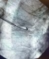
Another from a few months ago, elderly, frail, unable to independently ambulate or transfer. Sent by spine surgeon, not a candidate for fusion due to medical comorbid, osteoporosis. 90% relief. I see this outcome routinely. Do the procedure right, on patient with the right indications, and they do well with only very rare exception. Then get them on an anabolic agent.

I’ve got hundreds more cases on my phone if you still think this procedure can’t be done properly in a consistent basis….
 

Attachments

  • IMG_8619.jpeg
    IMG_8619.jpeg
    311.6 KB · Views: 134
  • IMG_8620.jpeg
    IMG_8620.jpeg
    453.3 KB · Views: 190
  • IMG_8621.jpeg
    IMG_8621.jpeg
    336.6 KB · Views: 155
  • IMG_8622.jpeg
    IMG_8622.jpeg
    316.3 KB · Views: 183
  • IMG_8623.jpeg
    IMG_8623.jpeg
    306 KB · Views: 114
  • IMG_8624.jpeg
    IMG_8624.jpeg
    297.5 KB · Views: 119
Yep. Absolutele ****e. Weird how a procedure doesn’t work when someone does it poorly. Here’s mine from yesterday with 100% relief. A little bursty. Waited till cement was very thick, inject small volume slowly, take breaks to harden and seal off cracks.
Beautiful…it’s pics like this that make me sometimes wanna get back into it
 
With the cement in the superior and inferior disc, we have no idea how much he tried to reposition and it continued to just spill out the fracture line. We don’t have true control ever in these cases. Are there nuances that some doctors know and others don’t? Yes. But we shouldn’t be judgmental, especially here where it looks like his bone filler was well placed midline vb/pedicle. The doctor who did the case is disappointed in the outcome, too. When/if you redo it, it will probably have a better result as there is some cement plugging the fracture line and the bone is more sclerotic now.
 
Advertisement - Members don't see this ad
With the cement in the superior and inferior disc, we have no idea how much he tried to reposition and it continued to just spill out the fracture line. We don’t have true control ever in these cases. Are there nuances that some doctors know and others don’t? Yes. But we shouldn’t be judgmental, especially here where it looks like his bone filler was well placed midline vb/pedicle. The doctor who did the case is disappointed in the outcome, too. When/if you redo it, it will probably have a better result as there is some cement plugging the fracture line and the bone is more sclerotic now.
You are right. I have had one really ugly looking leak that I could not get to stop despite adjustments and waiting in a highly compressed and comminuted fracture. I would be embarrassed if it was posted publicly.

Since then, if it is highly compressed with large voids in the endplate and not really room to adjust bone filler tip much ie high risk for leak, I have switched to a curved bone filler to allow for more adjustments in tip location, i.e. not just following the single straight path of entry. Even when it is not highly compressed, I have found just waiting to be unsuccessful at resolving the start of a leak. I don’t hesitate to re-stylet and adjust tip and bone filler location. If dense/sclerotic bone with minimal balloon expansion, I go to the curette to create some channels /voids to lower the pressure of cement injection. I have not found “egg-shelling” to be too helpful. I also do not think Osteopearl would help me in these highly compressed more complex fractures in the 2 cases I posted
 
Screenshot 2026-03-17 065851.png


Acute neck pain on right. H/o C3-7 ACDF 3 years ago. Hodgkin's Lymphoma, Osteomyelitis T10-11 in 2016.
WBC 4
ESR/CRP mildly elevated.
MRI with STIR edema at this joint and in soft tissues.
Not toxic appearance, afebrile.
 
Last edited:
Advertisement - Members don't see this ad
No disc. Ankylosing spondylitis with a bit of OPLL. No surgery needed. Neuro intact. Fused C2-S1 from disease. C spine OPLL not fully appreciated but MRI C and T spine next week. Got Radiology on board to do addendum on CT above.
 
No disc. Ankylosing spondylitis with a bit of OPLL. No surgery needed. Neuro intact. Fused C2-S1 from disease. C spine OPLL not fully appreciated but MRI C and T spine next week. Got Radiology on board to do addendum on CT above.
Stuff like this is why I keep coming back to work. Not bc of all the shiny new toys…that typically don’t work well
 
View attachment 417314
Didn’t know inflammatory markers peak after spine surgery and don’t return to baseline for 4 weeks. Good to know. Thanks
 
Advertisement - Members don't see this ad
I love a good cleft fill. Had two today.The t7 collapsed a lot between MRI three weeks ago and today. Lots of air in the bone with the balloon lifting as much as I could safely.
 

Attachments

  • IMG_0096.jpeg
    IMG_0096.jpeg
    338.1 KB · Views: 100
  • IMG_0093.jpeg
    IMG_0093.jpeg
    231.8 KB · Views: 93
  • IMG_0094.jpeg
    IMG_0094.jpeg
    268.7 KB · Views: 118
  • IMG_0095.jpeg
    IMG_0095.jpeg
    325.6 KB · Views: 93
  • IMG_0085.jpeg
    IMG_0085.jpeg
    290.3 KB · Views: 66
  • IMG_0088.jpeg
    IMG_0088.jpeg
    263.3 KB · Views: 99
  • IMG_0089.jpeg
    IMG_0089.jpeg
    215.4 KB · Views: 108
  • IMG_0090.jpeg
    IMG_0090.jpeg
    248.8 KB · Views: 98
  • IMG_0091.jpeg
    IMG_0091.jpeg
    93 KB · Views: 111
View attachment 418111

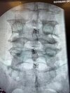
Revisiting this image. MVA 30 years ago. Patient reports Fx and experimental treatment. Markers in body, facet, and spinous process. WTH is this?
Per Claude

Those are almost certainly tantalum bead markers from Roentgen Stereophotogrammetric Analysis (RSA), a research imaging technique developed by Selvik at Lund University in the 1970s and used heavily in spine trials from the mid-1990s through mid-2000s to measure sub-millimeter intervertebral motion. The 0.5–1.0 mm beads were impacted into cancellous bone with a spring-loaded injector through small pilot holes — no plates, screws, or hardware tracts — which is why they remain as isolated radiodense dots in body, facet, and spinous process decades later. Given a ~1996 MVA and “experimental treatment,” this patient was most likely enrolled in a lumbar fusion cage or early dynamic stabilization trial of that era.
 
Advertisement - Members don't see this ad
Rare one. Baastrups. Prior neg mbb.
 

Attachments

  • IMG_0450.jpeg
    IMG_0450.jpeg
    287.7 KB · Views: 85
  • IMG_0451.jpeg
    IMG_0451.jpeg
    337.9 KB · Views: 86
  • IMG_0460.jpeg
    IMG_0460.jpeg
    420.9 KB · Views: 88
  • IMG_0461.jpeg
    IMG_0461.jpeg
    382.5 KB · Views: 86
  • IMG_0452.jpeg
    IMG_0452.jpeg
    297 KB · Views: 90
  • IMG_0453.jpeg
    IMG_0453.jpeg
    318.2 KB · Views: 98
  • IMG_0454.jpeg
    IMG_0454.jpeg
    329.6 KB · Views: 85
  • IMG_0462.jpeg
    IMG_0462.jpeg
    286.1 KB · Views: 88