
You are using an out of date browser. It may not display this or other websites correctly.
You should upgrade or use an alternative browser.
You should upgrade or use an alternative browser.
Pictures of the Week
Started by Epidural

Get help with your application
Use all the free resources available to you from SDN: articles, guides, expert advising, forums discussions, and school research.
Hard to see needle tip on lat but I'm guessing not quite there yet. CLO is so much better.I’ve gotten this pattern a few times in my career. What’s happening here
View attachment 419262
View attachment 419263

May be delaminating and pocketing contrast between layers. Try to aspirate it back out and make a slight adjustment-probably a bit deeper, but would make that decision in CLO.I’ve gotten this pattern a few times in my career. What’s happening here
View attachment 419262
View attachment 419263
Advertisement - Members don't see this ad
My guess: Too shallow but it’s spread anterolateral along the lamina, so in lateral it is superimposed on the epidural space. In this case I think an ipsilateral oblique would show you more clearly what’s going on.I’ve gotten this pattern a few times in my career. What’s happening here
View attachment 419262
View attachment 419263

You know I swear I took a CLO. Only reason I get a lateral is if the CLO looks weird. I must’ve forgot to save it and it had to have shown the needle anterior to the lamina otherwise I’d would’ve just advanced. 🤔My guess: Too shallow but it’s spread anterolateral along the lamina, so in lateral it is superimposed on the epidural space. In this case I think an ipsilateral oblique would show you more clearly what’s going on.
not there yet. blobogram. external spread.
why i dont like using a lot of contrast to advance, so i dilute it out by mixing contrast with saline - about 1:3 contrast to saline - in the LOR syringe. then when i think im in, i switch to full contrast syringe with tubing.
why i dont like using a lot of contrast to advance, so i dilute it out by mixing contrast with saline - about 1:3 contrast to saline - in the LOR syringe. then when i think im in, i switch to full contrast syringe with tubing.
Not that I was aware ofHard to say with the penetration but any signs of bifida occulta there on the pre-op images?

Came in for shoulder injection under fluoroscopy. Battery pack external. Pump internal. Icd and barostim present.
Did injection with patient standing and leaning on table.
Another endoscopic case. L3/4 left side with an L4 radic - transforaminal / SAP approach, utilized a high speed burr, thinned out the bone, removed the overgrown yellow ligament and the herniated disc in the foramen and lateral recess. U can see the traversing L4 nerve root in these progression pics at the bottom - great pulsations. Re-assessed today 4 weeks post op - pain free. LFG
Attachments

FIFYAnother endoscopic case. L3/4 left side with an L4 radic - transforaminal / SAP approach, utilized a high speed burr, thinned out the bone, removed the overgrown yellow ligament and the herniated disc in the foramen and lateral recess. U wish you can see the traversing L4 nerve root in these progression pics at the bottom - great pulsations. Re-assessed today 4 weeks post op - pain free. LFG
Awesome work.
FIFY
Awesome work.
you can see - idk why it autocorrected to wish
Haha. I did that. I can’t see anything. I’d love to have you teach me.you can see - idk why it autocorrected to wish

Yea, I can see less there than when mom came home with the ultrasound pics for my baby brother in 1995.
Could get her in for a ganglion impar if needed but she will try a
Sheepskin toast coach first
Continue Lyrica
Continue stretching exercises
Dragon dictate did this.
Sheepskin toast coach first
Continue Lyrica
Continue stretching exercises
Dragon dictate did this.
Advertisement - Members don't see this ad
me googling this and got:Could get her in for a ganglion impar if needed but she will try a
Sheepskin toast coach first
but we dont need EBM. just take lobels word.
know any good inexpensive versions to resell as DME?

Sheepskin Car-Cush® 13
Same great Car-Cush ® product with the added luxury of soft genuine Sheepskin! Orthopedic seat cushion designed to prevent and relieve back pain.
 tushcush.com


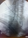

bad ass - nice caseCervical rf with deep brain stim. Wires on same side of rf, can see at far left on ap. Used bipolar to be extra cautious.
You using 18 or 20g?Cervical rf with deep brain stim. Wires on same side of rf, can see at far left on ap. Used bipolar to be extra cautious.
Normally 18. 20 on this one though.You using 18 or 20g?
Another kypho case with a large cleft, my favorite. Always very satisfying to see that thing fill up so densely. Just about always a home run outcome as well, including this one, 100% relief pod1. Used curved system for access and curved bone filler to minimize risk of leak.
Attachments


Is that a CLO? Would you mind talking through what you are looking for in that view? I typically use AP/Lat for cervical RFA but lower facets are always a nightmare.Cervical rf with deep brain stim. Wires on same side of rf, can see at far left on ap. Used bipolar to be extra cautious.
Yeah. Every single case. Makes these very easy.Is that a CLO? Would you mind talking through what you are looking for in that view? I typically use AP/Lat for cervical RFA but lower facets are always a nightmare.
Modified swimmers for MBB as well
Also, lots of good older threads on the forum from years ago.
Shows you the foramen and ensures that your tip is away from it. Also shows that you're on bone on a second angle (AP and foraminal view).Is that a CLO? Would you mind talking through what you are looking for in that view? I typically use AP/Lat for cervical RFA but lower facets are always a nightmare.
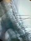
Had another good CLO case that may be helpful for you. C5-7, no visualization of 6 or 7 in lateral. Keys are:Is that a CLO? Would you mind talking through what you are looking for in that view? I typically use AP/Lat for cervical RFA but lower facets are always a nightmare.
1-ideally see your top needle on a lateral view so you can confirm the level, depth, and how high/low on pillar, as you don’t get the latter aspect as well on clo.
2- caudal tilt in clo until you see joint lines, so you now have similar information to lateral view.
Attachments


I have the local anesthetic for genicular RFA figured out. The key is to put the c arm in lateral and take a local needle from lateral to medial to the area you target and drop in 1-2ml .5% ropi. It takes less than a minute to do all 3. Then you go to AP, 1% lido for the skin and as deep as the local needle will go. Place your rf needles. 2% lido through the rf needles. I then get the probes out and let the MA test motors. I know it isn’t needed but we are just killing time and they aren’t capable of knowing when or when not to test motors. Then lesion. They went from crying out to chilling after I added the initial ropi. It probably has 8 minutes before you start ablating to work.
Blast from the past but….just did this approach (ropi first in lateral) for the first time with a patient who’s had genic RFA elsewhere and so had an expectation it would be rough. He also didn’t have a driver so couldn’t give him a PO benzo.
The guy was stunned when I told him we were 1/2 way through the first burn. Couldn’t feel a thing. I was stunned too lol.
Tip of the hat to @BobBarker
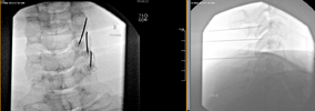

final needle placement looks ok, but i prefer to come more medial to lateral -- less chances of getting in trouble that wayCervical RFA feedback appreciated
Advertisement - Members don't see this ad

Cervical RFA feedback appreciated
Attachments
Missed target at C4 bilaterally. C4 runs in the middle to upper 1/3rd of the trapezoid, not bottom 1/3rd. Right C4 needle also too posteriorCervical RFA feedback appreciated
2 column to 3 column. Decompression and kypho.
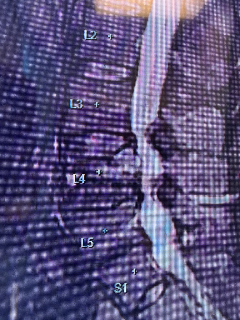
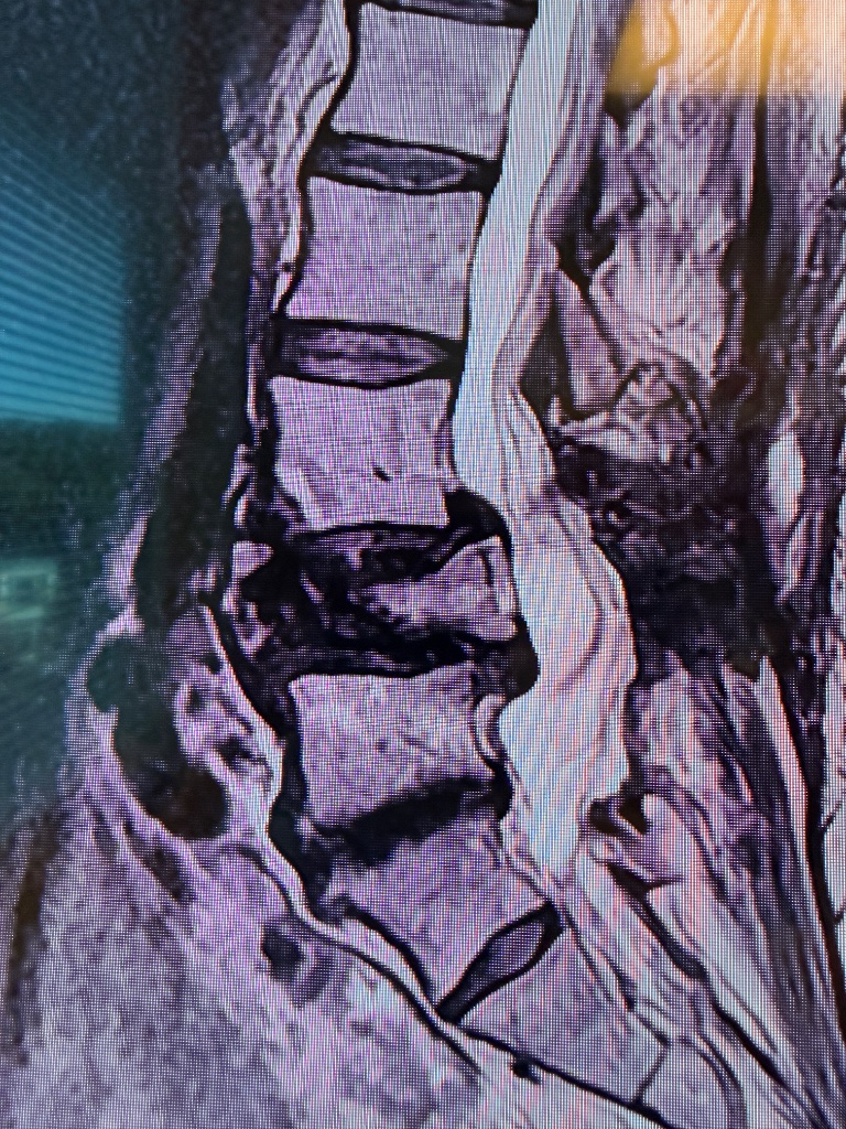
1 year ago is first pic. 1 week ago is new MRI. Patient underwent decompression and kypho.
Ongoing pain. Seeing as new pt tomorrow.
1 year ago is first pic. 1 week ago is new MRI. Patient underwent decompression and kypho.
Ongoing pain. Seeing as new pt tomorrow.
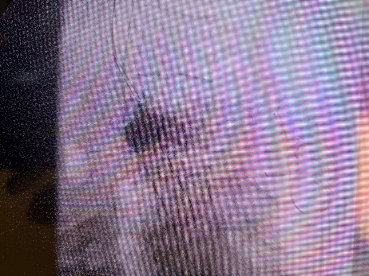
I was asked to do a c2 tfesi…. I’ve done plenty of c1-2 joints over the years and several c2 ganglion in fellowship. Used that approach, prone posterior. Contrast initially in the joint. Pulled back slightly, repeated contrast with DSA.
Attachments




Thanks, agreed I did put some of the injectate in the joint.Solid work man.
Any reason not to do 3-4mg of dex in the joint and then inject the remaining into the epidural space? Stenosis there is usually spondolytic right? Ever tried the facet overflow technique?
I haven’t done that, but seen it done before at joints below, not 1-2 though
solid work, what was the indication? I recently got a referral from rads for C1-2 block followed by C2 and C3 root block but MRI looks pristine. Hes insistentI was asked to do a c2 tfesi…. I’ve done plenty of c1-2 joints over the years and several c2 ganglion in fellowship. Used that approach, prone posterior. Contrast initially in the joint. Pulled back slightly, repeated contrast with DSA.
Thanks. This was directly referred to me by spine surgeon. has C2 root compression from the hypertrophic c1-2 lateral articulation.solid work, what was the indication? I recently got a referral from rads for C1-2 block followed by C2 and C3 root block but MRI looks pristine. Hes insistent
Higher risk procedure on a pristine MRI… No dice from me. Maybe mbb if whiplash, but I wouldn’t do a c1-2 or ctfesi on normal mri

Don’t want to lose a referral source but I would not do that.solid work, what was the indication? I recently got a referral from rads for C1-2 block followed by C2 and C3 root block but MRI looks pristine. Hes insistent
Don’t want to lose a referral source but I would not do that.
I agree 100%. If the referral is from radiology, why don’t they have IR do it? Also, it sounds like it isn’t even indicated anyway.
Sorry it was from neurology seeing them for headaches. My autocorrect charged to radsI agree 100%. If the referral is from radiology, why don’t they have IR do it? Also, it sounds like it isn’t even indicated anyway.
Just do occipital NB.Sorry it was from neurology seeing them for headaches. My autocorrect charged to rads
A neurologist trying to dictate what procedures we do is actually probably more ridiculous than radiology trying to.
If the neurologist is concerned about cervicogenic headache then I think third occipital nerve, C3 and C4 diagnostic medial branch blocks would be reasonable.
If the neurologist is concerned about cervicogenic headache then I think third occipital nerve, C3 and C4 diagnostic medial branch blocks would be reasonable.
2 column to 3 column. Decompression and kypho.
View attachment 422446View attachment 422447View attachment 422448
1 year ago is first pic. 1 week ago is new MRI. Patient underwent decompression and kypho.
Ongoing pain. Seeing as new pt tomorrow.
Advertisement - Members don't see this ad
Was a burst fx. Immediate post op above but poor pic. Had decompression and kypho by NS. And now this. Guessing corpectomy and fusion 2 levels above and below.Is that a poorly intercalated cement blob that pushed out and a posterior fragment that retropulsed in? It's like there's a reason some would have fused across it...
So, are you goina BVNA it?
A little atrophy.
Similar threads


- Replies
- 29
- Views
- 1K

- Replies
- 33
- Views
- 2K
- Replies
- 30
- Views
- 5K
