Step 1 Complicated Concepts Thread

This forum made possible through the generous support of SDN members, donors, and sponsors. Thank you.

TheSeanieB

Full Member
10+ Year Member
Joined
Dec 17, 2010
Messages
263
Reaction score
1
ASK AND ANSWER TOUGH QUESTIONS RELATED TO STEP 1.

Starting with me:
physiologic chloride shift - When CO2 diffuses into a RBC, it quickly converts with H2O to H+ and HCO3- so that CO2 will continue to passively diffuse into the RBC. The HCO3- is then excreted into the plasma by a Cl-/HCO3- exchanger. When the RBC enters the pulmonary capillaries, the process reverses. HCO3- is taken up by exchange for a Cl-. It combines with H+ to creates CO2 +H2O. The CO2 then diffuses out of the RBC and ultimately into the alveoli. This process allows for maximal CO2 excretion by a RBC.

Members don't see this ad.
 
Last edited:
  • Like
Reactions: 1 user
To add a little to the chloride shift- The HCO3- can diffuse out of the cell, but the H+ cant. If you didnt have the chloride shift a potential would build up so to keep the rbc at its normal membrane potential chloride shifts inside while hco3- diffuses out. HCO3- is more soluble in blood, and it can buffer acids, while the H+ inside can shift the oxygen affinity curve to the right enabling hb to unload o2 more readily.

Difficult concept-- pelvic anatomy. lol not really a concept, but thats what im focusing on right now.
 
For me:

lac operon
acid/base stuff, especially when it involves arrows
heart sounds
some genetics stuff
 
For me the countercurrent flow thing in the nephron. I haven't been able to understand it since high school ...
 
Members don't see this ad :)
Was having issues with brainstem anatomy/lesions

found this guy, have heard of it/seen it posted before just thought I would share again for those who havent seen it. 20 mins actually reading it and some practice questions was better then our whole Cranial Nerve section of Neuro.
http://lifeinthefastlane.com/2009/05/brainstem-rules-of-4/

Thanks for this. Will be reading over that soon.

Can we turn this thread into the "Official 2013 Step I Study Period Questions Thread"? I'm seeing a lot of separate, one question threads asking about a lot of misunderstood concepts, etc., and I propose we just ask the questions on this thread and have them answered here instead of starting an entirely new thread for every single question. Just a suggestion. It might make things easier.

i second this idea...
 
i second this idea...

We'll see if this works... I'll start.

In a previous thread, someone was talking about IgM vs. IgG antibody production in vaccines against polysaccharide antigen only and conjugated vaccines, respectively. I understand that the protein is necessary for presentation to the T-cells and the whole IgG class switching bit, but what about the unconjugated vaccine and how it induces immunity?

I thought IgM antibodies were nonspecific and only produced in acute infections, so how do unconjugated vaccines provide immunity against specific strains?
 
I thought IgM antibodies were nonspecific and only produced in acute infections, so how do unconjugated vaccines provide immunity against specific strains?

IgM antibodies do have a limited amount of specificity generated by VJ/VDJ rearrangement. Without somatic hypermutation (T-Cell required for this), you just get low-affinity antibodies.
 
We'll see if this works... I'll start.

In a previous thread, someone was talking about IgM vs. IgG antibody production in vaccines against polysaccharide antigen only and conjugated vaccines, respectively. I understand that the protein is necessary for presentation to the T-cells and the whole IgG class switching bit, but what about the unconjugated vaccine and how it induces immunity?

I thought IgM antibodies were nonspecific and only produced in acute infections, so how do unconjugated vaccines provide immunity against specific strains?

IgM has specificity, but doesn't have all the advantages of IgG (opsonization, placental crossing) so it's not the best immunoglobulin. Just because it's the first Ig doesn't mean it's solely present during the acute phase. An unconjugated pneumovax lasts 5ish years.
 
Was having issues with brainstem anatomy/lesions

found this guy, have heard of it/seen it posted before just thought I would share again for those who havent seen it. 20 mins actually reading it and some practice questions was better then our whole Cranial Nerve section of Neuro.
http://lifeinthefastlane.com/2009/05/brainstem-rules-of-4/

You got anymore stuff like this? This was simply amazing. Honestly, it just saved me having to go thru kaplan neuro
 
Hey does anyone have a good image that can show me the location of subepithelial deposits versus subendothelial deposits in kidney diseases
 
Members don't see this ad :)
thx, but even looking at these images I can't tell the difference between subepi and subendo. Can you help explain it to me w/ picture examples?

I was never able to understand this at all. I'm convinced it's just made up.

Sent from my SAMSUNG-SGH-I717
 
thx, but even looking at these images I can't tell the difference between subepi and subendo. Can you help explain it to me w/ picture examples?

Here you go.

dTbFkE8.png
 
Here you go.

dTbFkE8.png

This is good for conceptualizing, so i appreciate it. I still need some EM pictures, so that I can have it down 100%.

I was never able to understand this at all. I'm convinced it's just made up.

Sent from my SAMSUNG-SGH-I717

Its a conspiracy brah.But since Goljanski emphasizes it so much in the audio I feel like I'm costing myself points by not knowing it. Did you have anything like that show up on your step?
 
Thanks for this. Will be reading over that soon.

Can we turn this thread into the "Official 2013 Step I Study Period Questions Thread"? I'm seeing a lot of separate, one question threads asking about a lot of misunderstood concepts, etc., and I propose we just ask the questions on this thread and have them answered here instead of starting an entirely new thread for every single question. Just a suggestion. It might make things easier.

this is a great idea!
 
This is good for conceptualizing, so i appreciate it. I still need some EM pictures, so that I can have it down 100%.


Its a conspiracy brah.But since Goljanski emphasizes it so much in the audio I feel like I'm costing myself points by not knowing it. Did you have anything like that show up on your step?

Nope.

Sent from my Nexus 7
 
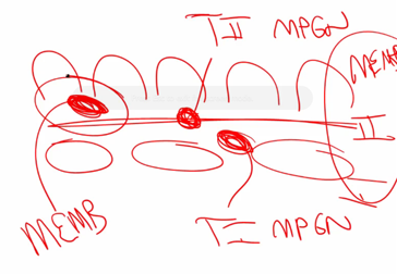
Podocyte foot process-----GBM------fenestrated glomerular endothelial cell
(I think technically all 3 together constitute the glomerular basemement membrane, but for conceptualizing consider the membrane between the two GBM)

Subepithelial deposits are between foot processes and the membrane.
da1c6ff17.png

You can tell that the deposits here are on the opposite side of the membrane than the RBCs (helps to tell where the endothelial side is). The GBM is the dark continuous street that circles around the picture continuously.

Subendothelial deposits are between the glom endothelial cell and the membrane.
RenalSlide23-1.png

You can see the enlongated endothelial cell nuclei with the subendothelial deposits on the same side of the GBM as the endothelium. On the opposite side of the GBM you can spot out the podocyte foot processes in a few areas. Also remember blood vessels make circles, so inside the circle is the endothelium, on the outside is the podocyte foot processes.


Intramembraneous deposit.
01em_36_wp.jpg
 
Last edited:
IgM antibodies do have a limited amount of specificity generated by VJ/VDJ rearrangement. Without somatic hypermutation (T-Cell required for this), you just get low-affinity antibodies.

IgM has specificity, but doesn't have all the advantages of IgG (opsonization, placental crossing) so it's not the best immunoglobulin. Just because it's the first Ig doesn't mean it's solely present during the acute phase. An unconjugated pneumovax lasts 5ish years.

Thanks, guys.
 
Podocyte foot process-----GBM------fenestrated glomerular endothelial cell
(I think technically all 3 together constitute the glomerular basemement membrane, but for conceptualizing consider the membrane between the two GBM)

Subepithelial deposits are between foot processes and the membrane.
da1c6ff17.png

You can tell that the deposits here are on the opposite side of the membrane than the RBCs (helps to tell where the endothelial side is). The GBM is the dark continuous street that circles around the picture continuously.

Subendothelial deposits are between the glom endothelial cell and the membrane.
RenalSlide23-1.png

You can see the enlongated endothelial cell nuclei with the subendothelial deposits on the same side of the GBM as the endothelium. On the opposite side of the GBM you can spot out the podocyte foot processes in a few areas. Also remember blood vessels make circles, so inside the circle is the endothelium, on the outside is the podocyte foot processes.


Intramembraneous deposit.
01em_36_wp.jpg

Thanks, this is helpful!
 
I am constantly getting the AIDs related CNS infections questions wrong. Anyone have a simple decision tree for these?

I know toxoplasmosis ->ring enhanced lesion, edema
JC virus ->visual and gait instabilities
HSV encephalitis->front lobe issues??
cryptococous meningitis?
CMV retinitis-doesn't this look like JC?

I had a question on USMLErx straight want me to tell the difference based on the patient having visual disturbances, lethargy, and gait instability... wtf.
 
Last edited:
I am constantly getting the AIDs related CNS infections questions wrong. Anyone have a simple decision tree for these?

I know toxoplasmosis ->ring enhanced lesion, edema
JC virus ->visual and gait instabilities
HSV encephalitis->front lobe issues??
cryptococous meningitis?
CMV retinitis-doesn't this look like JC?

I had a question on USMLErx straight want me to tell the difference based on the patient having visual disturbances, lethargy, and gain instability... wtf.

I'm starting to realize more and more than epidemiology matters a lot on some of those questions. I've caught myself being able to rule out specific organisms or tumors based on how old the patients were. Maybe they gave you other clues that you might have missed. Then again, it's Rx. I hate Rx, but Phloston is a huge advocate so I'm going to try to soldier through as much as I can.
 
I'm starting to realize more and more than epidemiology matters a lot on some of those questions. I've caught myself being able to rule out specific organisms or tumors based on how old the patients were. Maybe they gave you other clues that you might have missed. Then again, it's Rx. I hate Rx, but Phloston is a huge advocate so I'm going to try to soldier through as much as I can.

yeah, I was hoping it was just a ****ty question? haha.
 
I am constantly getting the AIDs related CNS infections questions wrong. Anyone have a simple decision tree for these?

I know toxoplasmosis ->ring enhanced lesion, edema
JC virus ->visual and gait instabilities
HSV encephalitis->front lobe issues??
cryptococous meningitis?
CMV retinitis-doesn't this look like JC?

I had a question on USMLErx straight want me to tell the difference based on the patient having visual disturbances, lethargy, and gain instability... wtf.

A lot of times you have to look at more than just the CNS issues to differentiate. CD4 count is big. Cryptococcus usually only causes meningitis.

A really high yield concept (imo) is HSV-1 is temporal lobe, for w/e reason HSV-1 loves itself some temporal lobe. HSV-2 would be the herpes of choice only in a neonate (thanks mom!).

Sorry I couldn't be more help I usually need a clinical scenario of some sort to be able to pick out the correct answer
 
I've been looking for a publication that lists and explain complicated concepts (not fully grasped from a review course) for step I with no luck. So, I would challenge, each person to define and explain a complicated concept that may be tested and is not well understood.

Starting with me:
physiologic chloride shift - When CO2 diffuses into a RBC, it quickly converts with H2O to H+ and HCO3- so that CO2 will continue to passively diffuse into the RBC. The HCO3- is then excreted into the plasma by a Cl-/HCO3- exchanger. When the RBC enters the pulmonary capillaries, the process reverses. HCO3- is taken up by exchange for a Cl-. It combines with H+ to creates CO2 +H2O. The CO2 then diffuses out of the RBC and ultimately into the alveoli. This process allows for maximal CO2 excretion by a RBC.

Can this explain why hyperchloremia causes a metabolic acidosis with a normal anion gap?
 
A lot of times you have to look at more than just the CNS issues to differentiate. CD4 count is big. Cryptococcus usually only causes meningitis.

A really high yield concept (imo) is HSV-1 is temporal lobe, for w/e reason HSV-1 loves itself some temporal lobe. HSV-2 would be the herpes of choice only in a neonate (thanks mom!).

Sorry I couldn't be more help I usually need a clinical scenario of some sort to be able to pick out the correct answer

no problem, this is great. any help I can get is awesome!
 
Difficult concept-- pelvic anatomy. lol not really a concept, but thats what im focusing on right now.

Does anyone know what we're expected to know with regards to pelvic anatomy? All those muscles are just confusing (or I've yet to find a source that actually teaches them simply and correctly).

Edit: to get seen
 
Last edited:
Can this explain why hyperchloremia causes a metabolic acidosis with a normal anion gap?

so...I did some research into hyperchloremic acidosis, especially why it is caused by saline infusion.

The answer is... confusing. Apparently, when you correct with the sodium bicarb the sodium is actually fixing the acidosis, not the bicarb. wtf? Maybe this review article is just wrong..

http://onlinelibrary.wiley.com/stor...8X.2003.tb00184.x.pdf?v=1&t=hgnqbeeg&f221f83c
 
Can this explain why hyperchloremia causes a metabolic acidosis with a normal anion gap?

I think by definition metabolic acidosis = low HCO3-. So if you happen to have high chloride in this situation it's called hyperchloremic (high Cl-) metabolic acidosis (low HCO3-) = normal gap-by the equation. I believe the two situations this occurs in is GI loss of base (diarrhea for example, Cl- still getting absorbed?) and renal tubular acidosis (can't absorb bicarb from urine, but you can absorb Cl-).

That's my best shot. I don't know if it's all absolutely factual but I thnk good enough for getting questions right .
 
Last edited:
Can this explain why hyperchloremia causes a metabolic acidosis with a normal anion gap?

I think it is best to associate hyperchloremic metabolic acidosis with acetazolamide toxicity aka decreased activity of PCT carbonic anhydrase (pg 525 FA 2012). If a patient is losing bicarb (decreased CA activity in nephron lumen) in the urine that will make them acidemic from the increased H+/HCO3- ratio in the blood.

Now, Cl- and HCO3 are antiported for one another in the PCT by the Base-/Cl- transporter (508 FA 2012). Since HCO3- is stuck in the tubular fluid it no longer antiports with Cl- in the PCT which leaves more Cl- in the bloodstream, hence, HYPERCHLOREMIA.

ANION GAP: The anion gap does not change because of Cl- and HCO3- inverse relationship (AG = Na - (Cl +HCO3)). If you lose HCO3- the subsequent increase in Cl- will balance out the anion gap as seen in the AG equation.

Hope that makes sense. Took me 20 min to figure out so I hope this shows up on my test haha. :laugh:
 
  • Like
Reactions: 1 user
I think by definition metabolic acidosis = low HCO3-. So if you happen to have high chloride in this situation it's called hyperchloremic (high Cl-) metabolic acidosis (low HCO3-) = normal gap. I believe the two situations this occurs in is GI loss of base (diarrhea, Cl- still getting absorbed?) and renal tubular acidosis (can't absorb bicarb from urine, but you can absorb Cl-).

That's my best shot. I don't know if it's all absolutely factual but I thnk good enough for getting questions right .

it also occurs in saline infusions.
 
I think it is best to associate hyperchloremic metabolic acidosis with acetazolamide toxicity aka decreased activity of PCT carbonic anhydrase (pg 525 FA 2012). If a patient is losing bicarb (decreased CA activity in nephron lumen) in the urine that will make them acidemic from the increased H+/HCO3- ratio in the blood.

Now, Cl- and HCO3 are antiported for one another in the PCT by the Base-/Cl- transporter (508 FA 2012). Since HCO3- is stuck in the tubular fluid it no longer antiports with Cl- in the PCT which leaves more Cl- in the bloodstream, hence, HYPERCHLOREMIA.

ANION GAP: The anion gap does not change because of Cl- and HCO3- inverse relationship (AG = Na - (Cl +HCO3)). If you lose HCO3- the subsequent increase in Cl- will balance out the anion gap as seen in the AG equation.

Hope that makes sense. Took me 20 min to figure out so I hope this shows up on my test haha. :laugh:
this is extremely helpful, thanks!
 
If you have non gap metabolic acidosis, don't you have to also be hyperchloremic? I thought that was in the definition-if you lose bicarb you have to balance that loss by gain of anion, either measurable (chloride) or unmeasurable (which would lead to an anion gap).
 
man, the crosssection of the spinal cord tracts SUCKS. It's way too simplified if you ask me.

better one:
http://upload.wikimedia.org/wikipedia/commons/b/b2/Spinal_cord_tracts_-_English.svg
I think you only really need to know the:

anterior + later corticospinal
dorsal columns(gracillus and cuneatus)
anterior + lateral spinothalamic

It seems like the spinocerebellar and extra-pyramidal(rubro,olivo, etc) are extraneous information that doesn't need to be committed to memory.
 
I just looked this up, it's solid.

Black = inhibit
Red = excite
Ignore the first arrow from the striatum to the D1 or D2. Just know that the striatum itself has D1, D2 receptors, and those will for the most part, selectively send out axons to the direct (D1 neurons) and indirect (D2 neurons) pathways. Just draw the first arrow from the striatum directly to GPi/SNr and directly to GPe to avoid confusion
 
Last edited:
http://www.vstretch.net
I just looked this up, it's solid.

Black = inhibit
Red = excite
Ignore the first arrow from the striatum to the D1 or D2. Just know that the striatum itself has D1, D2 receptors, and those will for the most part, selectively send out axons to the direct (D1 neurons) and indirect (D2 neurons) pathways. Just draw the first arrow from the striatum directly to GPi/SNr and directly to GPe to avoid confusion[/QUOTE]

Pretty good picture. I photoshopped it a bit for clarity.

[IMG]http://i.imgur.com/7rrGoZj.png
 
This thread is a great idea. Ok so I have a question about the coagulation cascade, specially factor VIII. So we all know heparin stimulates anti thrombin III which degrades 2, 7,9,10,11 and 12. Sooo not 8??? Is that because factor 8 is "protected" by vWF or is there some mechanism I am missing. Also why not follow PTT and PT with someone on heparin? Both the intrinsic and extrinsic pathways are inhibited.
 
This thread is a great idea. Ok so I have a question about the coagulation cascade, specially factor VIII. So we all know heparin stimulates anti thrombin III which degrades 2, 7,9,10,11 and 12. Sooo not 8??? Is that because factor 8 is "protected" by vWF or is there some mechanism I am missing. Also why not follow PTT and PT with someone on heparin? Both the intrinsic and extrinsic pathways are inhibited.

Indeed, they both share a common pathway. Here is an explanation from an old thread: http://forums.studentdoctor.net/archive/index.php/t-954944.html


It just happens that the difference in PTT vs PT elevation in heparin is much higher than the reverse difference for warfarin. That is to say, you need pretty big doses of heparin to see a meaningful rise in PT. You don't need huge doses of warfarin to see a rise in PTT. I think almost any presentation of heparin wrt PT and PTT will show a normal PT and increased PTT. The reverse is not true of warfarin therapy (you might see elevated PT and PTT, although PT (INR) is the preferred tracking metric).

and you can see the test goal values are very different: http://surgery.about.com/od/beforesurgery/qt/PTPTTINRtests.htm
Prothrombin Time Blood Test-PT

This test is done to evaluate the blood for its ability to clot. It is often done before surgery to evaluate how likely the patient is to have a bleeding or clotting problem during or after surgery.

Normal PT Values: 10-12 seconds (this can vary slightly from lab to lab)

Common causes of a prolonged PT include vitamin K deficiency, hormones drugs including hormone replacements and oral contraceptives, disseminated intravascular coagulation (a serious clotting problem that requires immediate intervention), liver disease, and the use of the anti-coagulant drug warfarin. Additionally, the PT result can be altered by a diet high in vitamin K, liver, green tea, dark green vegetables and soybeans.

Partial Thromboplastin Time Blood Test-PTT

This test is performed primarily to determine if heparin (blood thinning) therapy is effective. It can also be used to detect the presence of a clotting disorder. It does not show the effects of drugs called “low molecular weight heparin” or most commonly by the brand name Lovenox.

Normal PTT Values: 30 to 45 seconds (this can value slightly from lab to lab)

Extended PTT times can be a result of anticoagulation therapy, liver problems, lupus and other diseases that result in poor clotting.
 
Are any of you guys using the rules of 4 for brainstem lesions????

It's basically this right:

the structure deficit tells you if the lesion is middle or lateral. The nerve deficit tells you how high or low in the brain stem it is. Together, you have the exact spot of the damage?
 
Are any of you guys using the rules of 4 for brainstem lesions????

It's basically this right:

the structure deficit tells you if the lesion is middle or lateral. The nerve deficit tells you how high or low in the brain stem it is. Together, you have the exact spot of the damage?

Pretty much, I actually like it and will make sure I have it into my head before test day. I've always just rote memorized up until now
 
Pretty much, I actually like it and will make sure I have it into my head before test day. I've always just rote memorized up until now

me too, I have never seen this before, it is legit awesome. I hated memorizing this stuff during my neuroscience block....

Is this a fair thing to say for blood supply?

medial medulla - ASA
lateral medulla - PICA
lateral pons - AICA

What does basilar get? medial pons + midbrain?
 
Top